Muscles of Back Deep Layers Biological Science Picture Directory
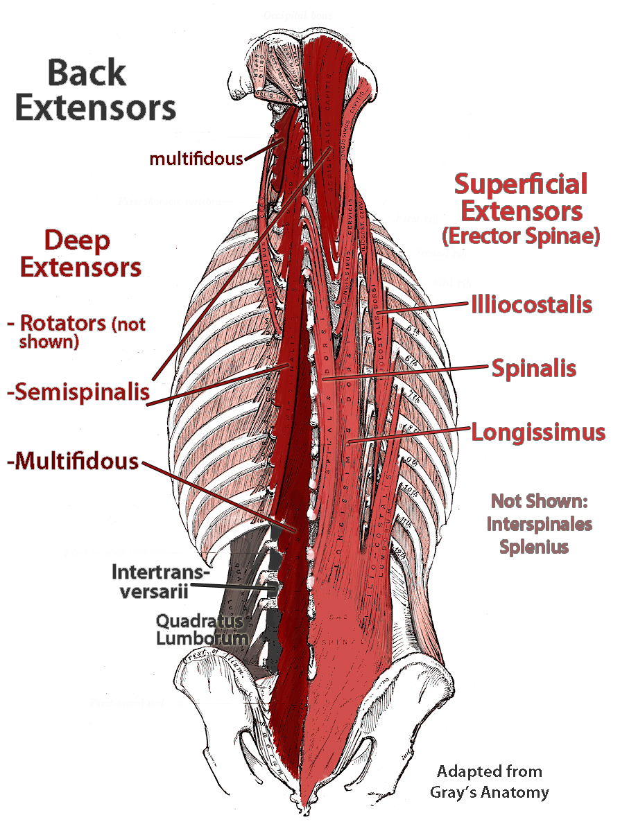
Erector spinae: three groups ("I long for spinach"), lateral to medial: illiocostalis: lateral. longissimus: in the middle. spinalis: medial. Transversospinales: three groups, from superficial to deep: semispinalis. multifidus. rotares. Learn all about the muscles of the back in this 3D video anatomy tutorial.

Anatomy of back muscles Diagram Quizlet
Back Muscles w/ pictures 18 terms Jenna_Carey1 Preview parts of the brain 33 terms kmd6gu Preview Muscle Labeling - Upper Leg 22 terms MonaL18 Preview Layers of the wall of the digestive tract 10 terms abreejtheriot Preview Muscle Labeling - Lower Leg

Muscles Labeled Front And Back Human Anatomy Body
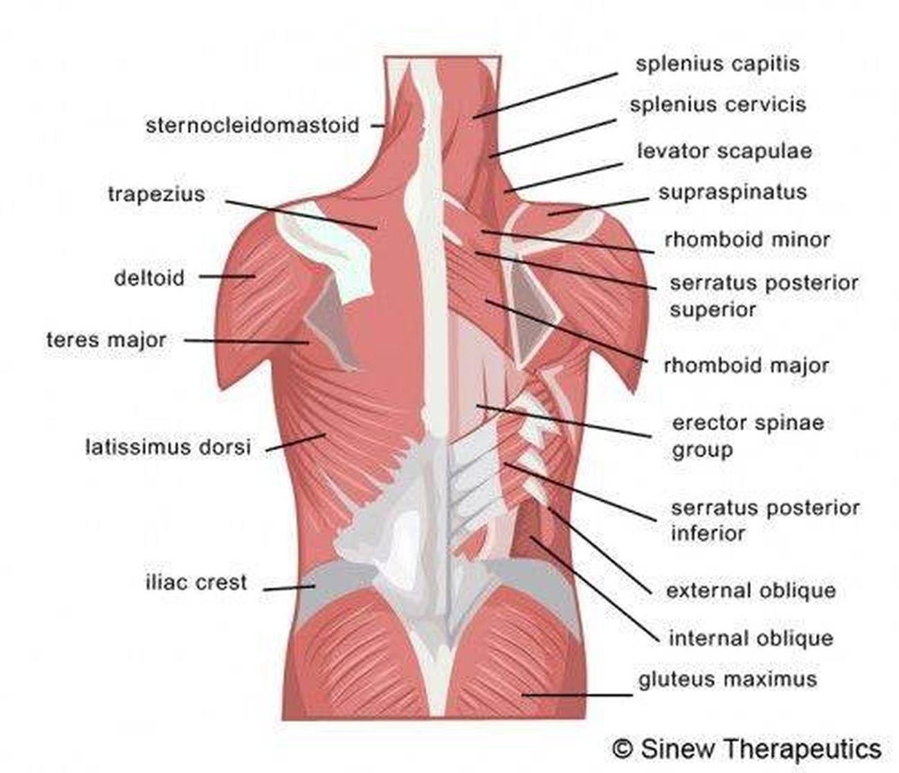
Figure \(\PageIndex{7}\): Muscles of the Neck and Back. The large, complex muscles of the neck and back move the head, shoulders, and vertebral column. (Image credit: "Muscles of the Neck and Back" by Openstax is licensed under CC BY 4.0) The erector spinae group forms the majority of the muscle mass of the back and it is the primary extensor.

Muscles of the Back TeachMeAnatomy
What are your back muscles? Your back has many different muscles. Some muscles support your spine and trunk. Others help you move your body, stand up straight and assist with breathing. Because your back muscles support so much of your weight and are responsible for so many movements, injuries to these muscles are common.

Back Muscle Diagram exatin.info
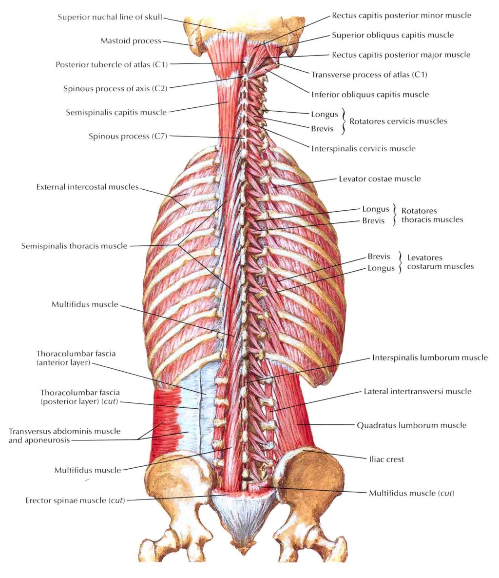
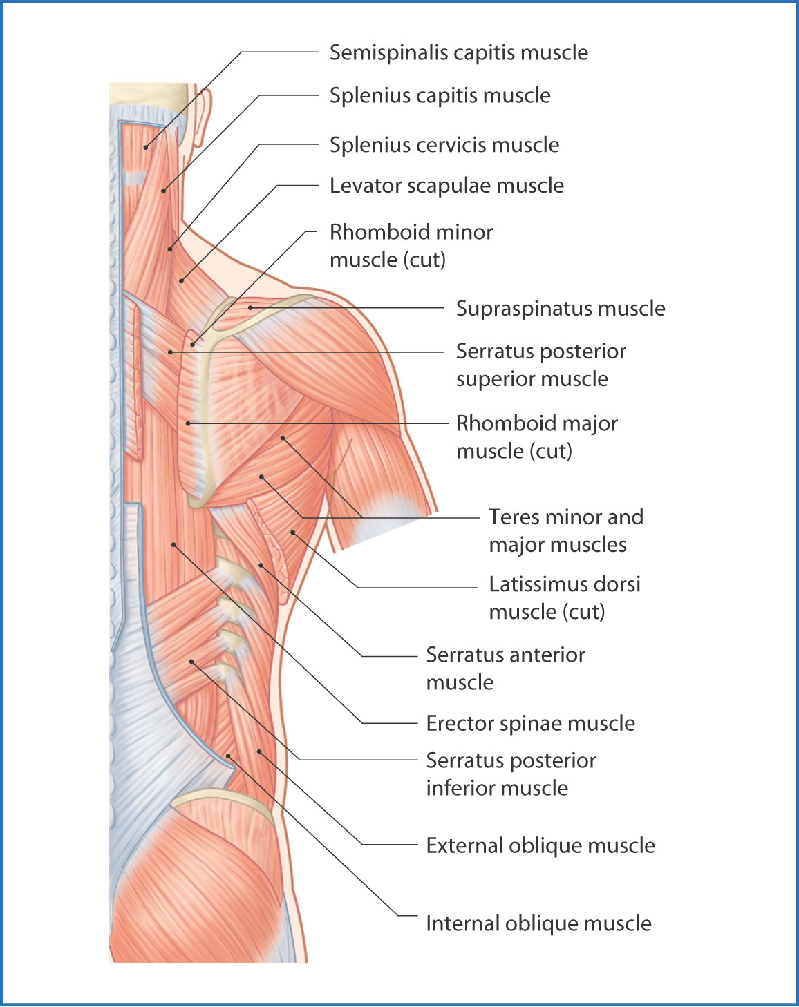
The deep back muscles, also called intrinsic or true back muscles, consist of four layers of muscles: superficial, intermediate, deep and deepest layers. These muscles lie on each side of the vertebral column, deep to the thoracolumbar fascia. They span the entire length of the vertebral column, extending from the cranium to the pelvis.
Pictures Of Back Muscles
Ligaments of the Back. 3D video tutorials and interactive modules on the anatomy of the back including anatomy of the musculature, vertebral column, joints and ligaments.
Pictures Of Back Muscles
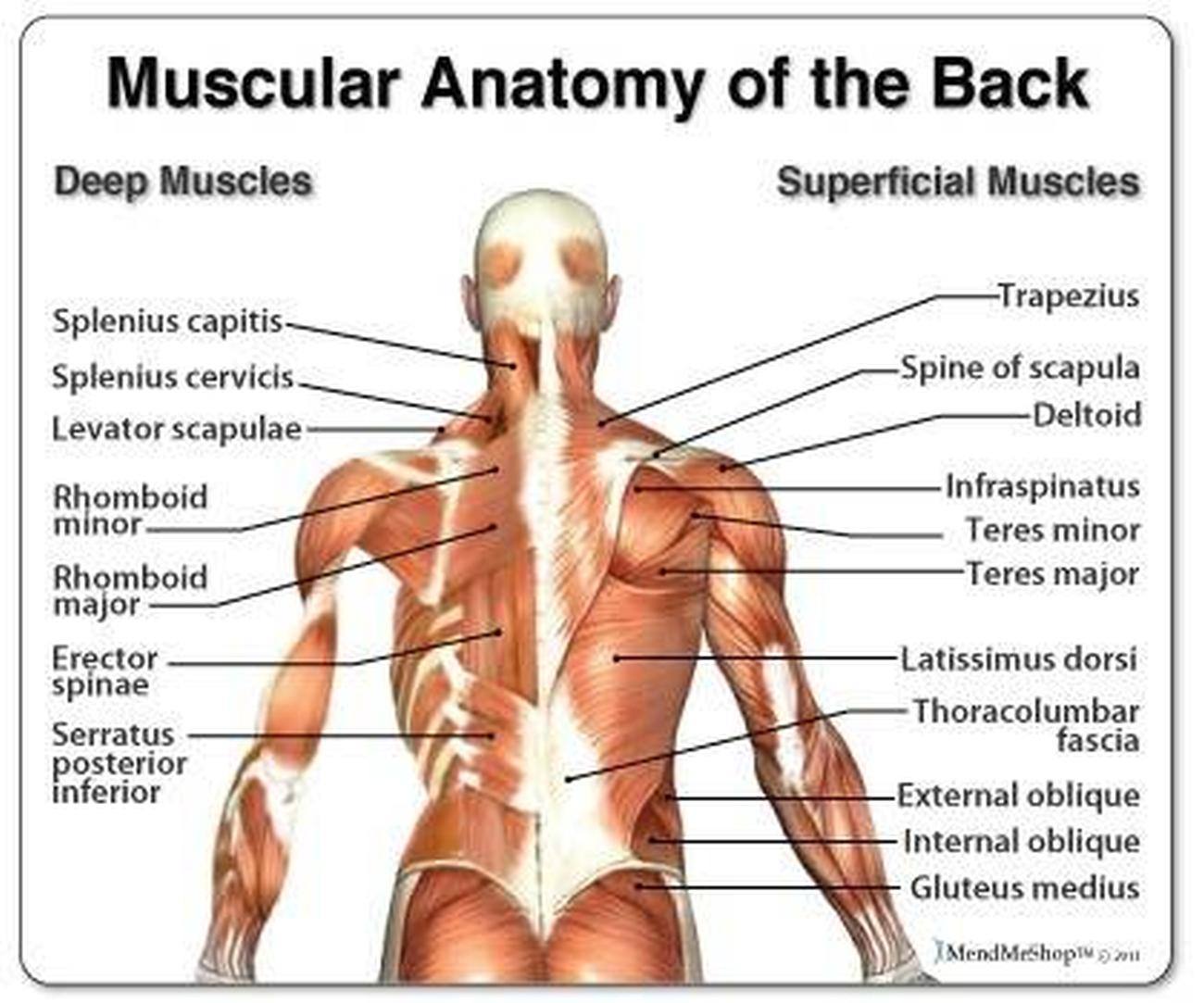
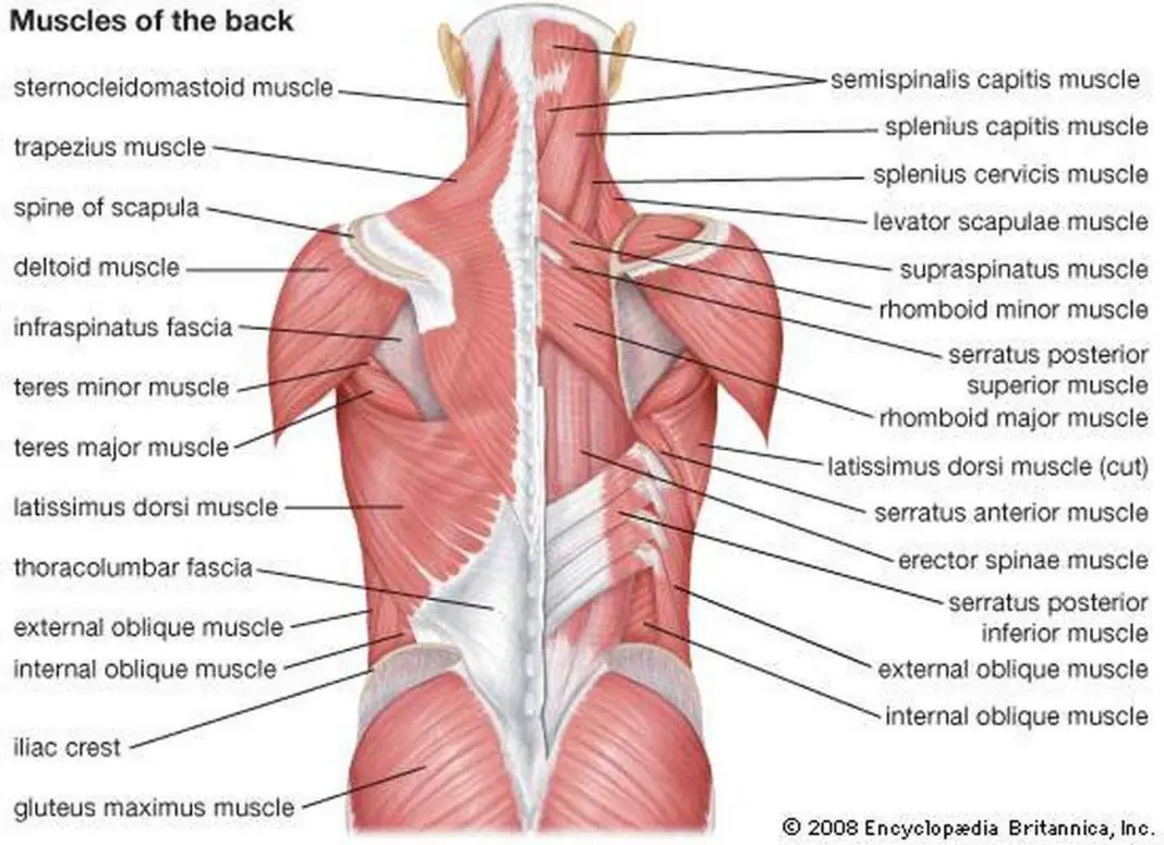
The muscles of the back categorize into three groups. The intrinsic or deep muscles are those muscles that fuse with the vertebral column. The second group is the superficial muscles, which help with shoulder and neck movements. The final group is the intermediate muscles, which help with the movement of the thoracic cage.

Diagram Of Hip.and Back.muscles qwlearn
The muscles of the back can be arranged into 3 categories based on their location: superficial back muscles, intermediate back muscles and intrinsic back muscles.The intrinsic muscles are named as such because their embryological development begins in the back, oppose to the superficial and intermediate back muscles which develop elsewhere and are therefore classed as extrinsic muscles.
Pictures Of Back Muscles
Muscles of the Back Image of the muscles of the back, labeled for reference and study.

Back Muscle Diagrams 101 Diagrams
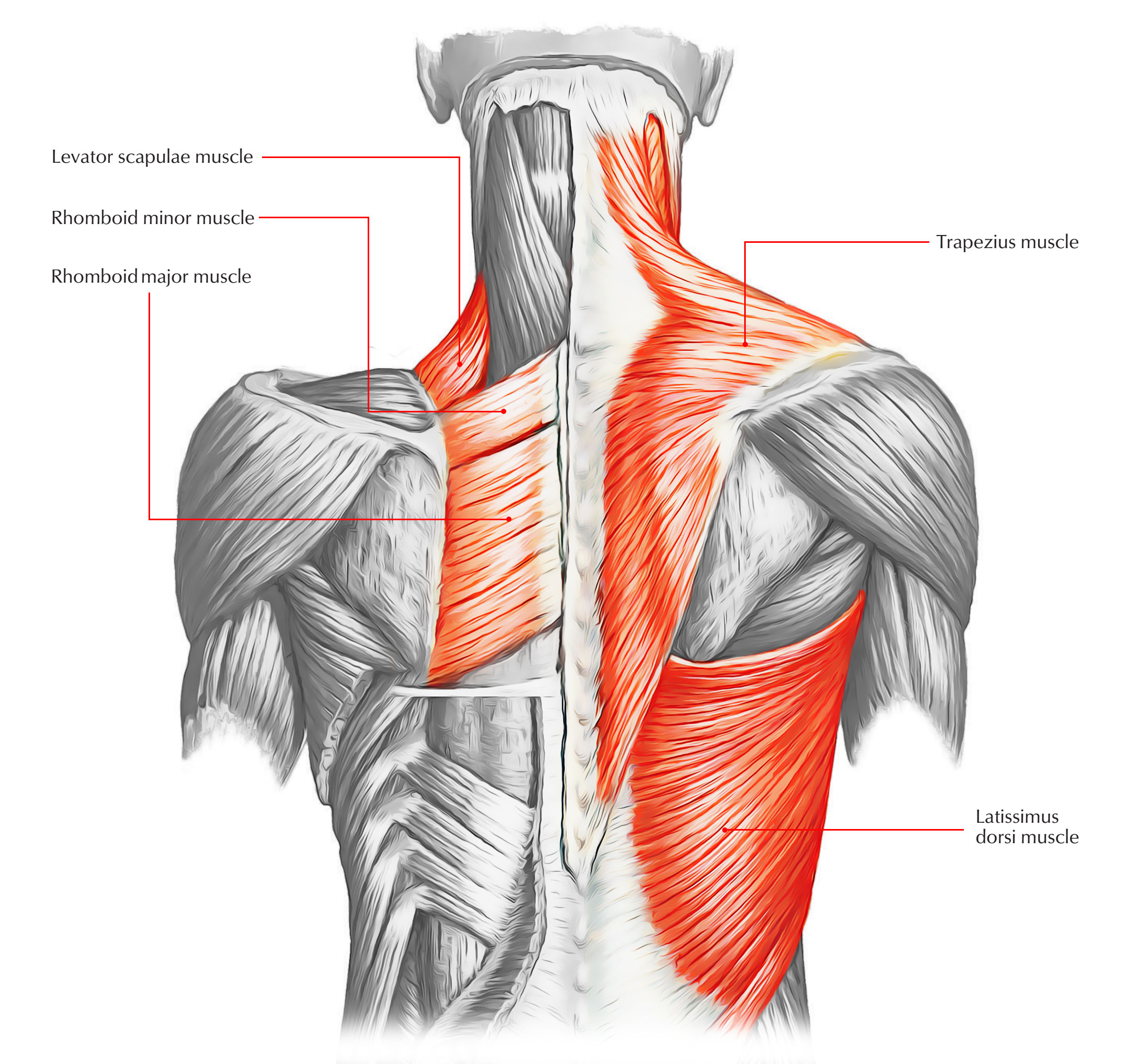
A back muscle that pulls the arm down and back. It is responsible for extension,adduction, and (medial) internal rotation of the shoulder joint. It also helps in extension and lateral flexion of the lumbar spine. The name means "widest of the back." This muscle supports the arm when it is moved above the head.
88+ Muscles Of The Back l2sanpiero
This online quiz is called Back muscles. It was created by member LilStride and has 13 questions.
Back Muscles Basicmedical Key
Anatomy of the back: spine and back muscles Author: Jana Vasković MD • Reviewer: Nicola McLaren MSc Last reviewed: November 03, 2023 Reading time: 14 minutes Recommended video: Superficial back muscles [17:28] Attachments, innervation and functions of the superficial muscles of the back. Back anatomy

Intrinsic Back Muscles Anatomy of the Torso Medical Library
Human body muscle diagrams Muscle diagrams are a great way to get an overview of all of the muscles within a body region. Studying these is an ideal first step before moving onto the more advanced practices of muscle labeling and quizzes. If you're looking for a speedy way to learn muscle anatomy, look no further than our anatomy crash courses .

Thoracic Mobility Impulse Chiro & Natural Therapies
The back is found posteriorly and includes the vertebral column, the muscles that support the back and the spinal cord. The vertebral column consists of 33 vertebrae which can be split up into 5 continuous sections. Each section is functionally different and is specialised for either weight-bearing, movement, protection and/or posture.

Back Muscles Diagram Quizlet
Study with Quizlet and memorize flashcards containing terms like Longissimus Capitis, Splenius Capitis, Serratus Posterior Superior and more.
deep muscles of lower back Biological Science Picture Directory
Latissimus Dorsi Your latissimus dorsi, or lats, are the largest individual muscles in your upper back. They run down the sides of your torso and, when developed through resistance training,.
