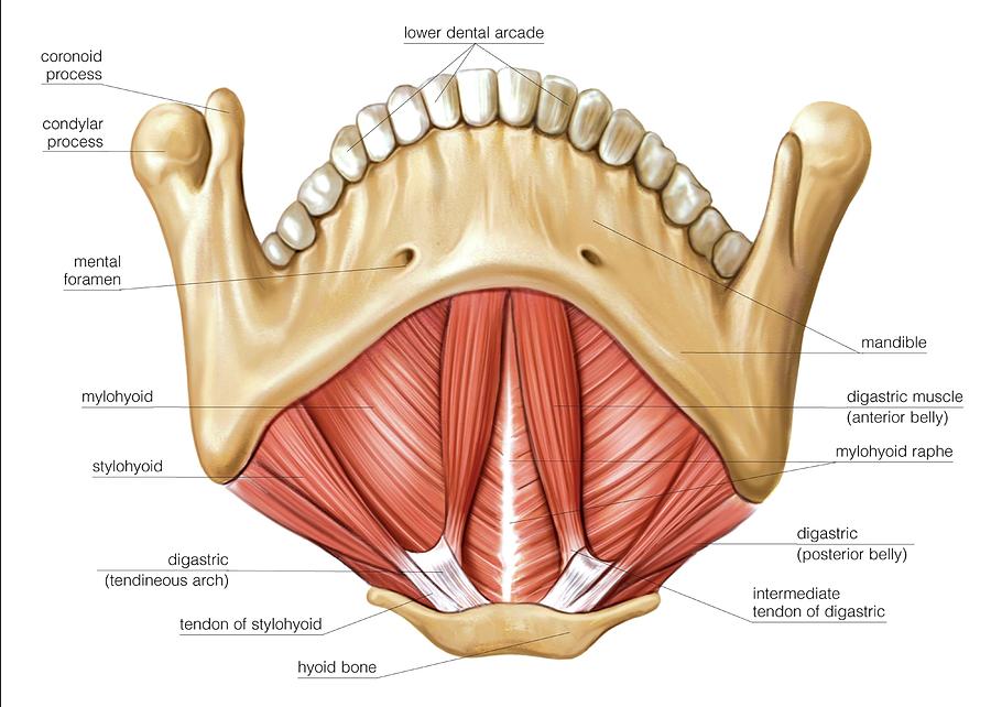
Muscles Of The Floor Of Mouth Photograph by Asklepios Medical Atlas
A wide range of pathologic processes may involve the floor of the mouth, the part of the oral cavity that is located beneath the tongue. They include lesions that arise uniquely in this location (eg, ranula, submandibular duct obstruction) as well as various malignancies, inflammatory processes, and vascular abnormalities that may also occur elsewhere in the head and neck.

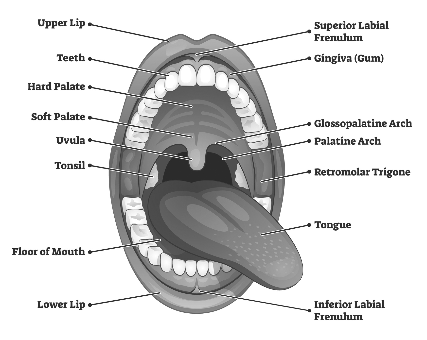
Schematic drawing of the oral cavity [97]. Download Scientific Diagram
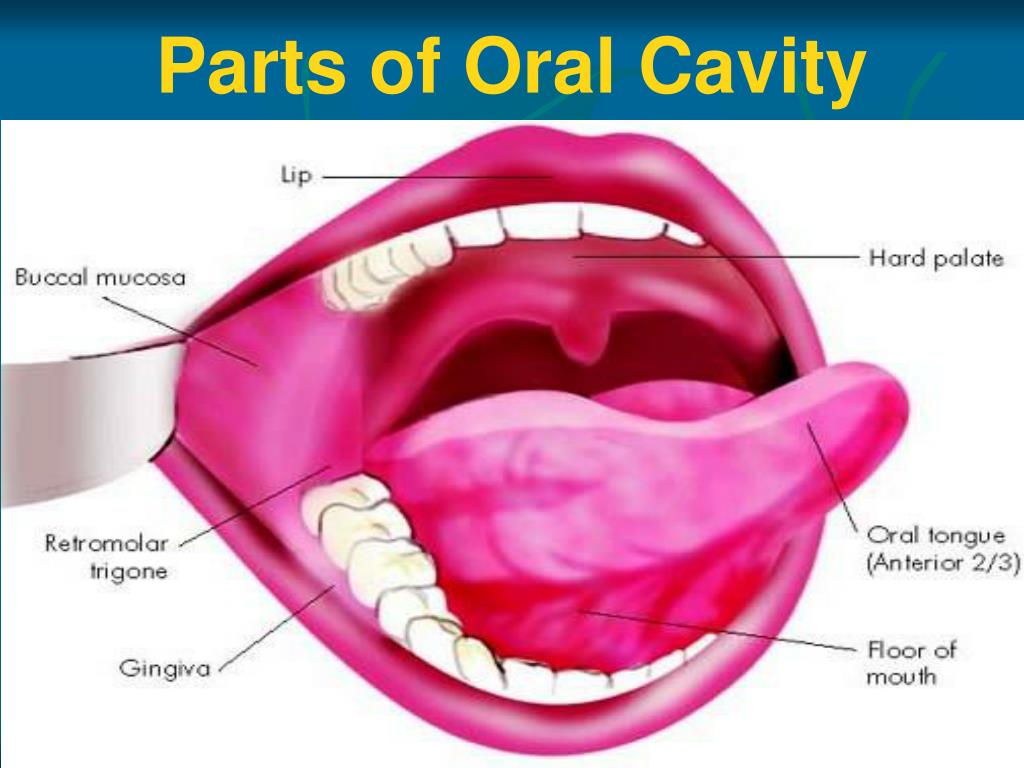
It consists of several different anatomically different aspects that work together effectively and efficiently to perform several functions. These aspects include the lips, tongue, palate, and teeth. Although a small compartment, the oral cavity is a unique and complex structure with several different nerves and blood vessels inside it.

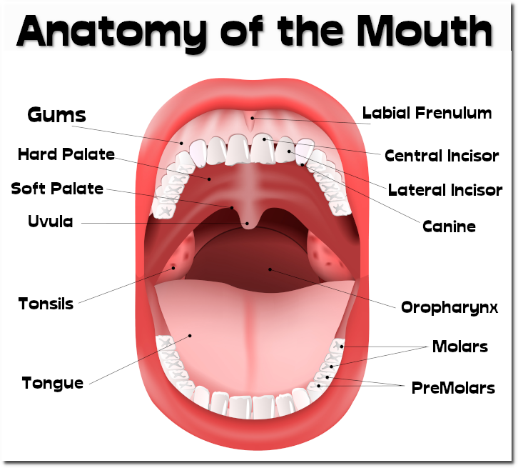
Detailed mouth anatomy
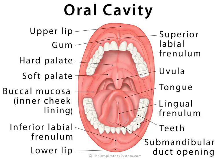
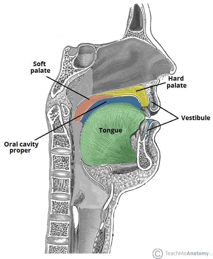
Structure Oral cavity Anatomy of the mouth. Floor of the mouth with lingual frenum and sublingual fold The mouth consists of two regions: the vestibule and the oral cavity proper. The vestibule is the area between the teeth, lips and cheeks. [3]
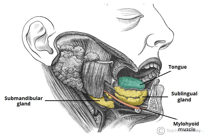
The Oral Cavity Divisions Innervation TeachMeAnatomy
The floor of mouth is an oral cavity subsite and is a common location of oral cavity squamous cell carcinoma . Gross anatomy The floor of mouth is a U-shaped space which extends (and includes) from the oral cavity mucosa superiorly, and the mylohyoid muscle sling 2,3 . Boundaries superiorly: oral mucosal space inferiorly: mylohyoid muscle 3
Floor Of Mouth Anatomy Pdf Free Bios Pics
Full text PDF Tools Share Abstract Familiarity with the radiologic anatomy and landmarks of the floor of the mouth is helpful for detecting and characterizing pathologic processes that occur there and extend to deep tissues and beyond.
Oral Floor Photograph by Asklepios Medical Atlas Pixels
The oral cavity spans between the oral fissure (anteriorly - the opening between the lips), and the oropharyngeal isthmus (posteriorly - the opening of the oropharynx). It is divided into two parts by the upper and lower dental arches (formed by the teeth and their bony scaffolding).
PPT Anatomy of Oral Cavity, Pharynx & Oesophagus PowerPoint
The mouth opens to the outside at the lips and empties into the throat at the rear; its boundaries are defined by the lips, cheeks, hard and soft palates, and glottis. It is divided into two sections: the vestibule, the area between the cheeks and the teeth, and the oral cavity proper.

Gross Anatomy Glossary Oral Cavity Draw It to Know It
A computed tomography (CT) technique is described which demonstrates the structures and tissue planes in the floor of mouth, tongue and oropharynx. The anatomy, which forms the basis for understanding pathological change, is given in detail and illustrated by axial and coronal images and line drawings.
What is the Oral Cavity
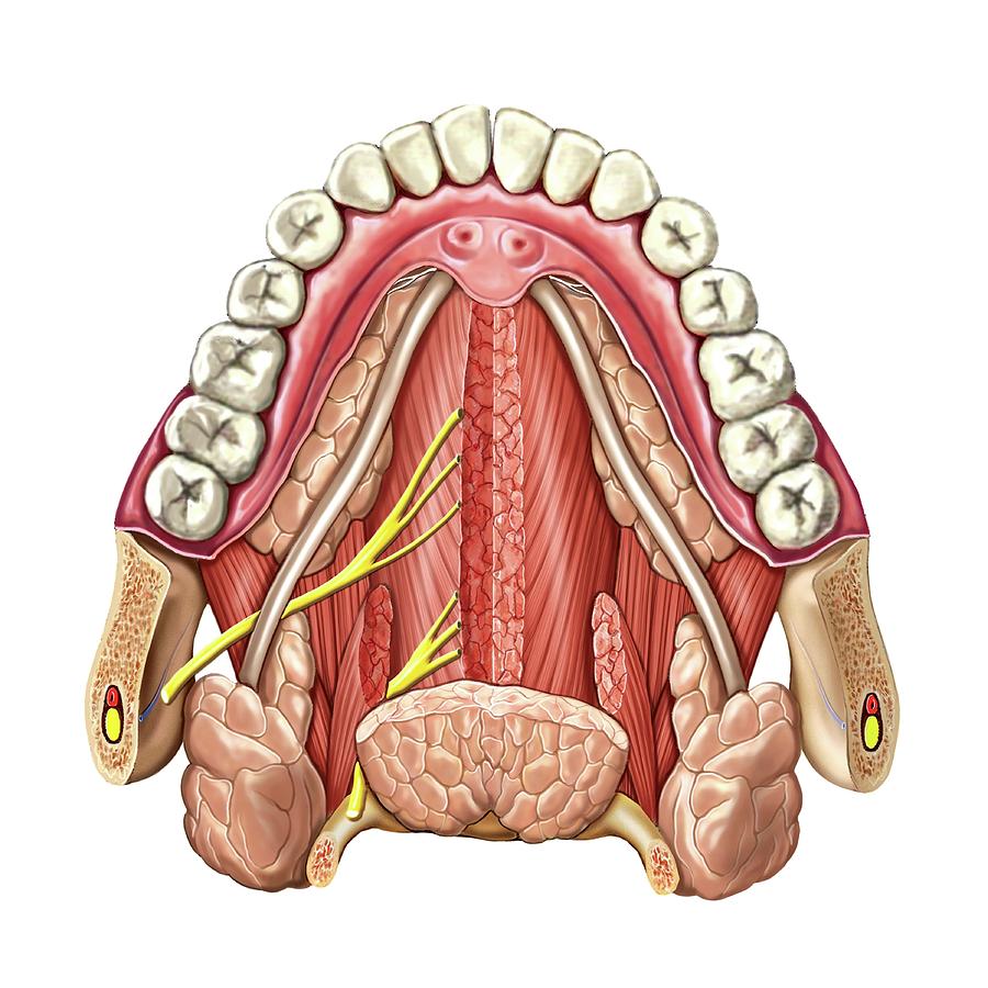
The floor of mouth (i.e., sublingual space) is a U-shaped region, bordered inferiorly by the mylohyoid muscle, laterally by the gingiva overlying the lingual surface of the mandible, superiorly by the oral tongue, and posteriorly at the insertion of the anterior tonsillar pillar into the tongue (Fig. 14-5). From: Oncologic Imaging, 2002

23.3 The Mouth, Pharynx, and Esophagus Anatomy & Physiology
Pros and Cons CT and MR are used for the majority of nondental imaging in the oral cavity. Plain radiographs, orthopantomography, and occlusal views remain useful tools for studying the teeth and mandible as discussed in Chapter 96.
The Oral Cavity Divisions Innervation TeachMeAnatomy
Anatomy of the Floor of the Mouth Fig. 1: Normal anatomy of the floor od the mouth on contrast enhanced MDCT studies. A, D - coronal, B - axial at the level of the mandible, E - axial at the level of the hyoid bone, C - coronal Important anatomical landmarks of the floor of the mouth - paired muscles and spaces:

25 The oral cavity and related structures Pocket Dentistry
What's my mouth's function? Your mouth supports many daily functions, including: Breathing. Talking. Chewing. Tasting. Swallowing. Eating. Drinking. Mouth function in digestive system Your mouth is where digestion begins. When you chew food, your salivary glands make saliva (spit). Saliva helps break down starches in the foods you eat.

AN3 08 Oral Cavity, Oropharynx, Swallowing StudyBlue
When we say 'mouth' we mean the oral cavity; a space in the lower part of the head that functions as the entrance to the digestive system. The content of the oral cavity determines its function. It houses the structures necessary for mastication and speech, which include the teeth, the tongue and associated structures such as the salivary glands.

PPT ORAL ANATOMY PowerPoint Presentation, free download ID2381675
(1) Department of Oral Surgery, Implant Surgery and Radiology, School of Dentistry, Faculty of Health Sciences, Aristotle University of Thessaloniki, Thessaloniki, Greece 8.1 General Anatomy and Ultrasonographic Features 8.2 Inflammatory Changes 8.2.1 Ranulas 8.3 Benign Tumors 8.3.1 Branchial Cysts 8.3.2 Thyroglossal Duct Cysts and Fistulas
Floor of the mouth
The anatomy of the tongue and floor of the mouth is readily discernible by computed tomography (CT) because of low-density fascial planes that outline the extrinsic musculature, lingual arteries, and hypoglossal nerves. Although the tongue is accessible to the examining finger, few patients can tolerate a detailed palpation. In planning for a partial glossectomy, CT scanning aids the surgeon.
Anatomy of the Mouth everythingherbs
Introduction. Radiological evaluation of the floor of the mouth (FOM), an anatomical compartment of the oral cavity, is complex and challenging. 1 The region harbours different types of tissues, including salivary glands, ducts, mucosa, submucosal soft tissues and the bony mandible and can be associated with a wide range of diseases, including congenital, inflammatory, infective, benign and.
