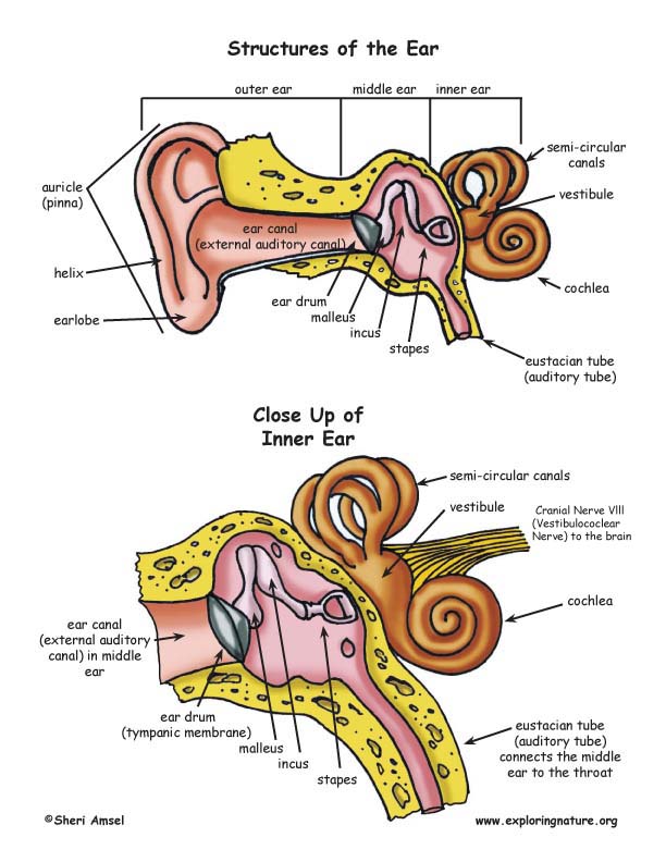
Structures of the Ear
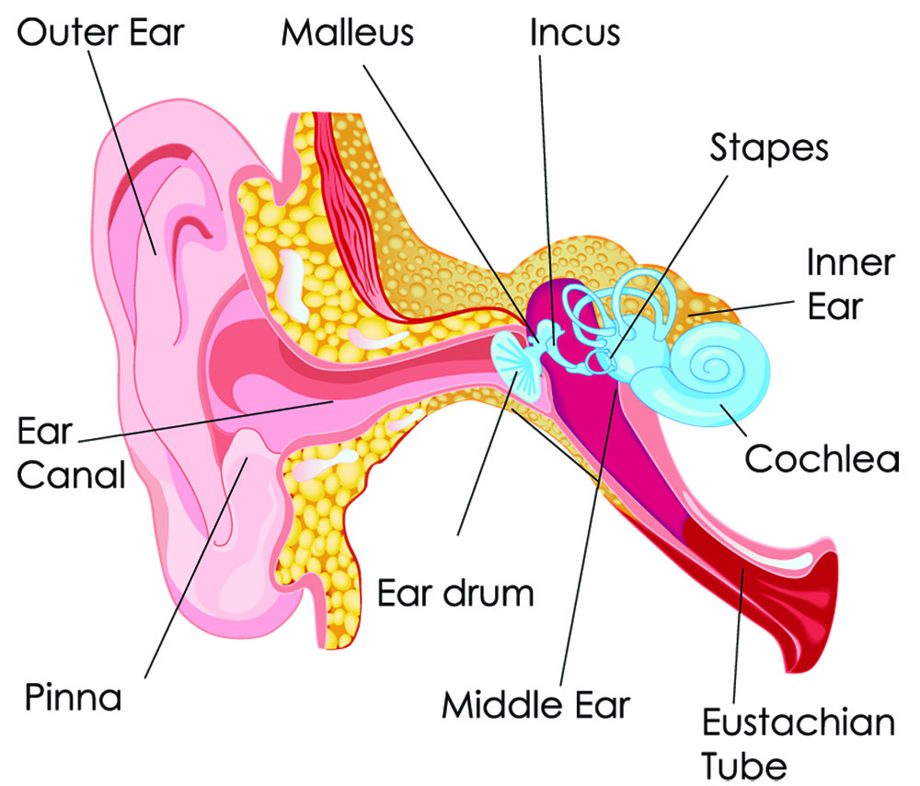
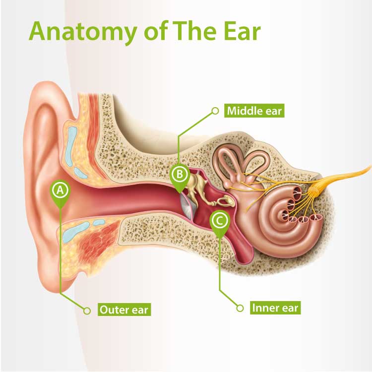
The ear is divided into three parts: Outer ear: The outer ear includes an ear canal that is is lined with hairs and glands that secrete wax. This part of the ear provides protection and.
[DIAGRAM] Inside Of Ear Diagram
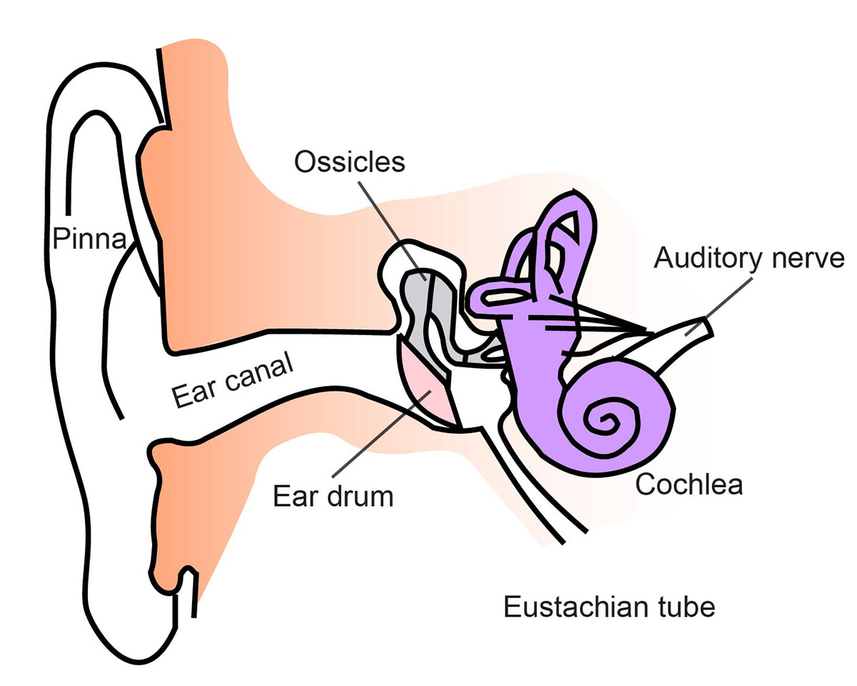
Here is a blank human ear diagram for you to label, so that you can memorize the different parts of this vitally necessary organ, for good.
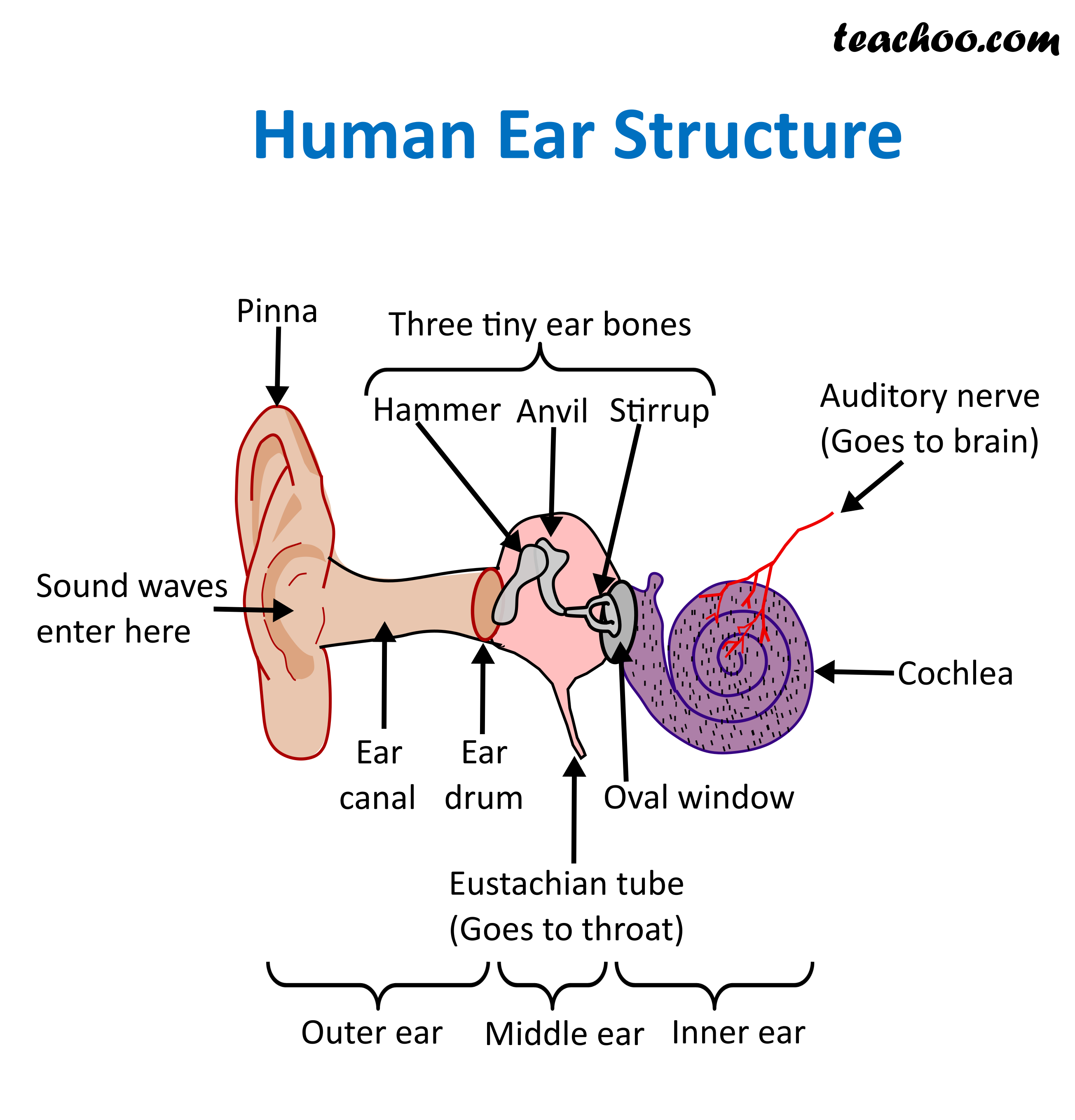
Structure and Function of Human Ear with Diagram Teachoo diagram
The purpose of the inner ear is to sense and process information about sound and balance, and send that information to the brain. Each part of the inner ear has a specific function. Cochlea: The cochlea is responsible for hearing. It is made up of several layers, with the Organ of Corti at the center.
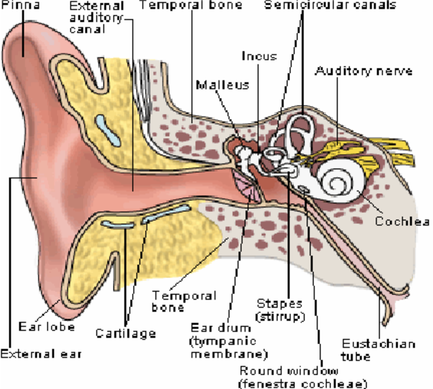
Anatomy of the Ear [4]. Download HighQuality Scientific Diagram
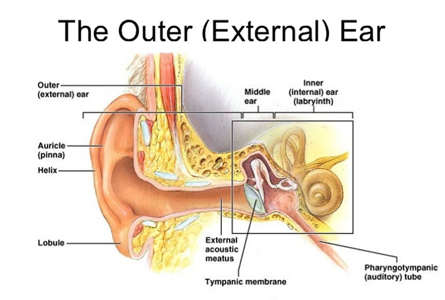
The medical term for the outer ear is the auricle or pinna. The outer ear is made up of cartilage and skin. There are three different parts to the outer ear; the tragus, helix and the lobule. The ear canal starts at the outer ear and ends at the ear drum. The canal is approximately an inch in length. The skin of the ear canal is very sensitive.

Structure of the human ear Ear anatomy, Human anatomy and physiology
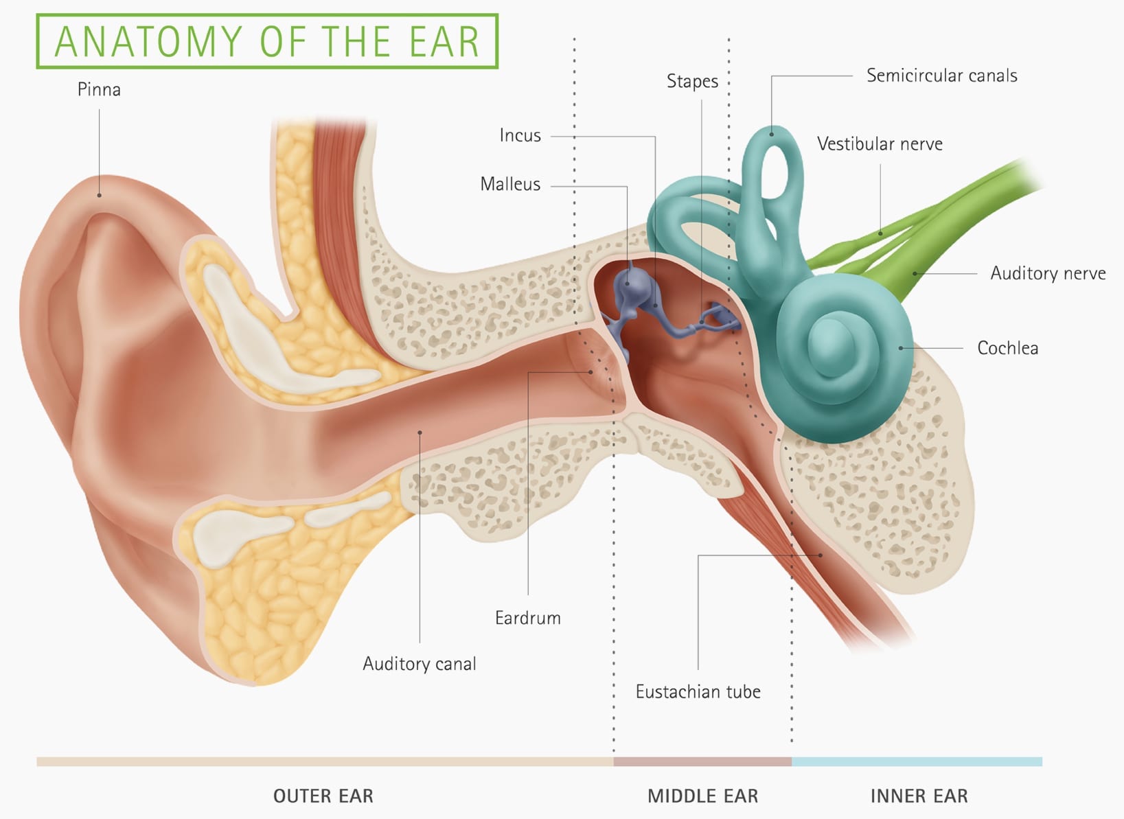
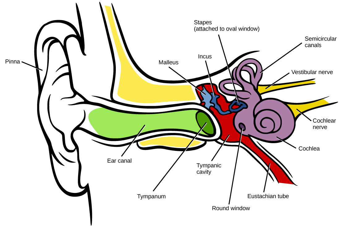
A brief description of the human ear along with a well-labelled diagram is given below for reference. Well-Labelled Diagram of Ear The External ear or the outer ear consists of Pinna/auricle is the outermost section of the ear. The external auditory canal links the exterior ear to the inner or the middle ear.

Labelling the EAR This is a one page worksheet that explores the
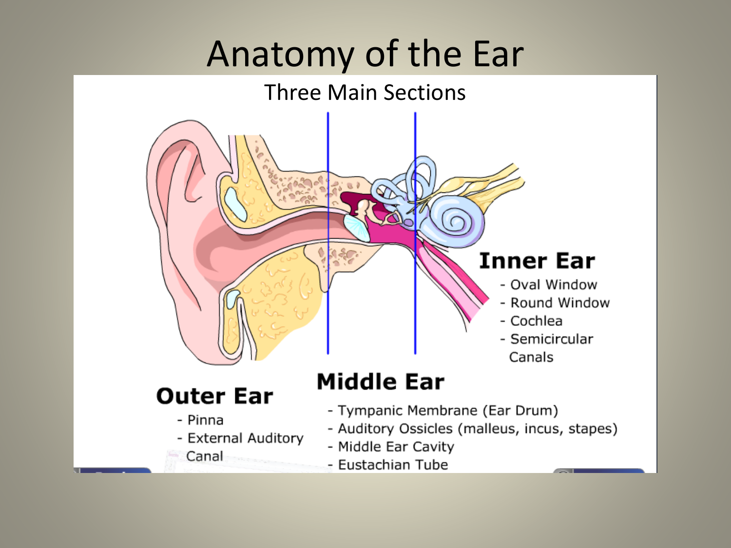
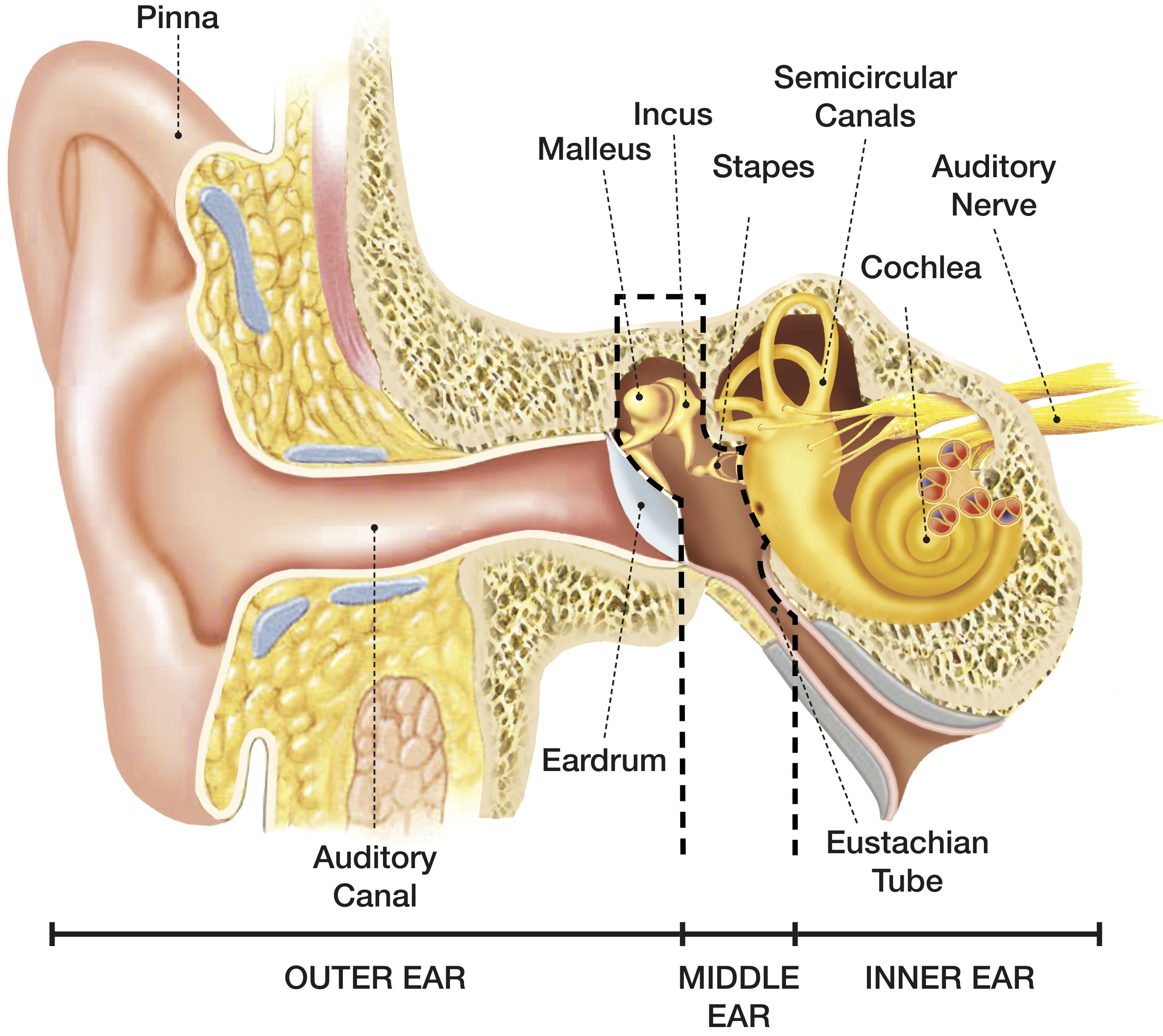
The ear diagram is one of the important topics for Class 10 and 12 students of the CBSE board and in this article, we will briefly explain the structure of the ear, its different parts and their functions. Parts of the Human Ear. The human ear consists of three different parts. These are: The outer ear. The middle ear. The inner ear
Ear Anatomy Chart
Inner Ear - Diagram and Description. The human ear comprises three parts, namely the external, middle and inner ear. The inner ear or labyrinth is the innermost part that consists of the bony and membranous labyrinth. The vestibular apparatus is a part of the inner ear that plays a vital role in maintaining equilibrium and posture.
Your Hearing Heritage Hearing
Chapter 1 - Introduction Manual Format How to examine the ears Suggested Procedure Chapter 2 - Testing Audiogram Tympanogram Chapter 3 - Ear Anatomy Ear Anatomy - Outer Ear Ear Anatomy - Inner Ear Ear Anatomy Schematics Ear Anatomy Images Chapter 4 - Fluid in the ear Fluid in the ear Discussion Fluid in the ear Outline Middle Ear Ventilation Tubes
Outer Ear Anatomy Outer Ear Infection & Pain Causes & Treatment
Protect your ears. If the noise is too loud, walk away, turn it down (Turn it to the Left), or use ear plugs. pinna ear canal ear drum hammer anvil stirrup Eustachian tube (connects to the nose) cochlea semicircular canals nerves (connect to the brain) Directions: Color in the diagram below using a different color for each part of the ear.
Ear Anatomy Diagram Images
Helix: The outermost curvature of the ear, extending from where the ear joins the head at the top to where it meets the lobule. The helix begins the funneling of sound waves into the ear; Fossa, superior crus, inferior crus, and antihelix: These sections make up the middle ridges and depressions of the outer ear. The superior crus is the first ridge that emerges moving in from the helix.
3 Things to Know About the Signal Path of the Auditory System — Pro
Download this blank ear diagram below Contents Ear anatomy overview Ear diagrams (labeled and unlabeled) Accelerate your learning with interactive quizzes Sources + Show all Ear anatomy overview Although it's not obvious to look at, the ear is anatomically divided into three portions: External (outer) ear Middle ear Inner ear
How We Hear Hearing Associates, Inc.
Your outer ear and middle ear are separated by your eardrum, and your inner ear houses the cochlea, vestibular nerve and semicircular canals (fluid-filled spaces involved in balance and hearing). What is the ear? Your ears are organs that detect and analyze sound. Located on each side of your head, they help with hearing and balance. Advertisement
How the ear works Triton Hearing
1/4 Synonyms: External auditory meatus, External acoustic pore , show more. The ear is a complex part of an even more complex sensory system. It is situated bilaterally on the human skull, at the same level as the nose. The main functions of the ear are, of course, hearing, as well as constantly maintaining balance.
Understanding how the ear works Hearing Link Services
The Ear; The Ear - Map Quiz Game. Cochlea; Ear canal; Eardrum; Eustachian tube; Incus; Inner ear; Malleus; Middle ear; Outer ear; Semicircular canals; Stapes; You need an account to play. Create challenge. 0/11 0 % 00:05 Click on Inner ear > Click on Inner ear. Game mode: Pin Type Show more game modes. Learn. Restart---
Parts Of The Ear Drawing at Explore collection of
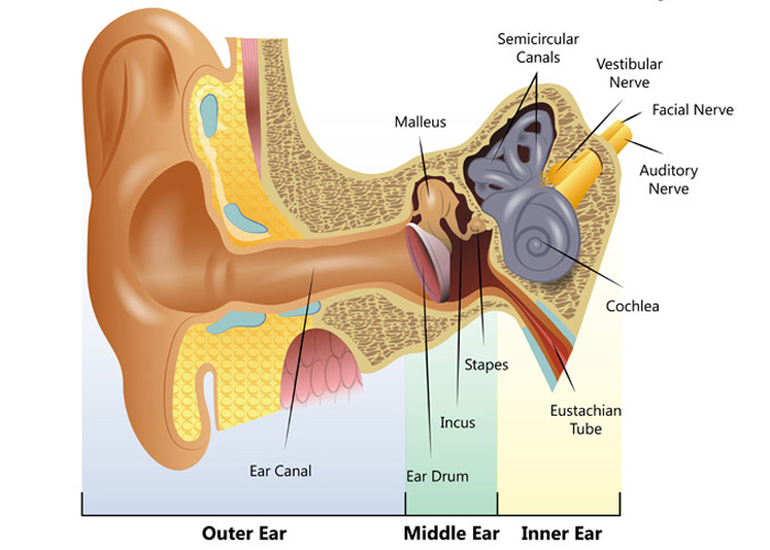
The Outer Ear When thinking about the anatomy of the ear, we should start with the outer ear, which is responsible for collecting sound waves from our environment and funneling them through the ear canal into the middle ear. The outer ear consists of the pinna (auricle) and the ear canal (meatus).

Human Ear Home Tuition Guwahati Assam Human ear diagram, Ear
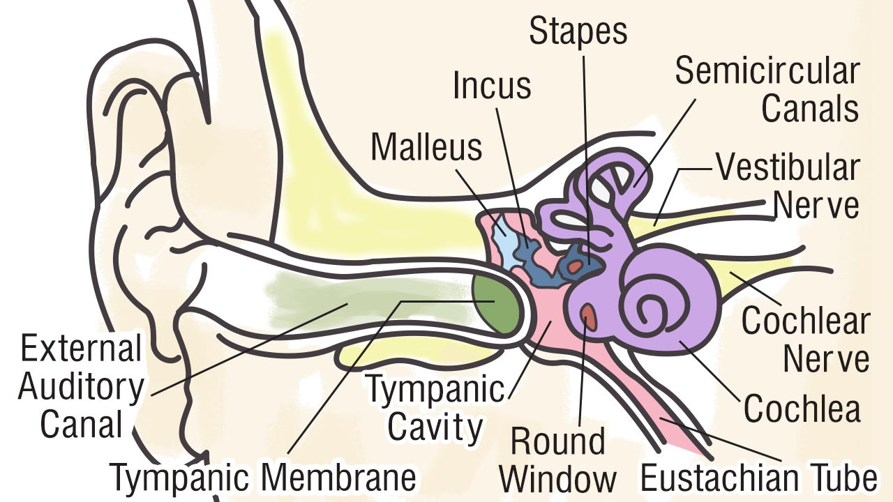
1: Diagram showing the structure of the human ear, detailing the parts of the outer, middle, and inner ear. Source publication +48 A Framework for Speechreading Acquisition Tools Thesis.
