PPT Anatomy of the Ear PowerPoint Presentation ID6102402
Özocak O, Unur E, Ülger H, Ekinci N, Aycan K, Acer N. Meatus acusticus internus'un morfometrisi ve varyasyonları. Erciyes Üniversitesi Sağlık Bilimleri Dergisi. 2004;13(3) 1-7. 4. Kobayashi H, Zusho H. Measurements of internal auditory meatus by polytomography. The British Journal of Radiology 1987; 60 (711): 209-14.
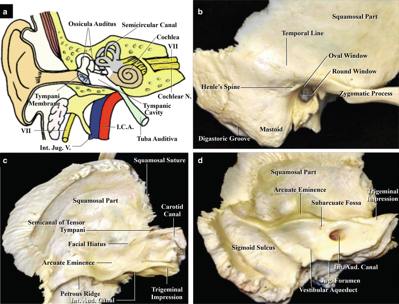
Temporal Bone Anatomy
Der Meatus acusticus internus ist ein kleiner, relativ kurzer Knochenkanal, der im Felsenbein (Pars petrosa ossis temporalis) verläuft. Er beginnt am Porus acusticus internus und endet an einer durchlöcherten Knochenplatte, die an das Innenohr grenzt.
external acoustic meatus bone location
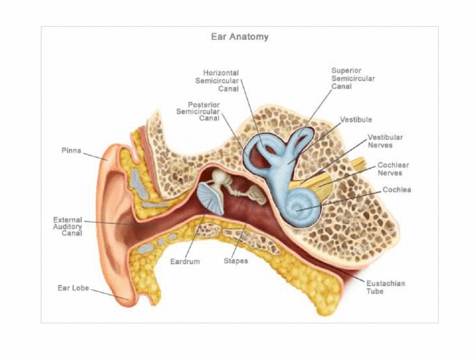
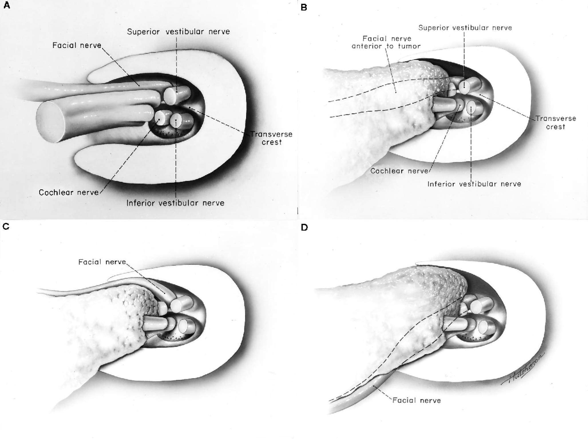
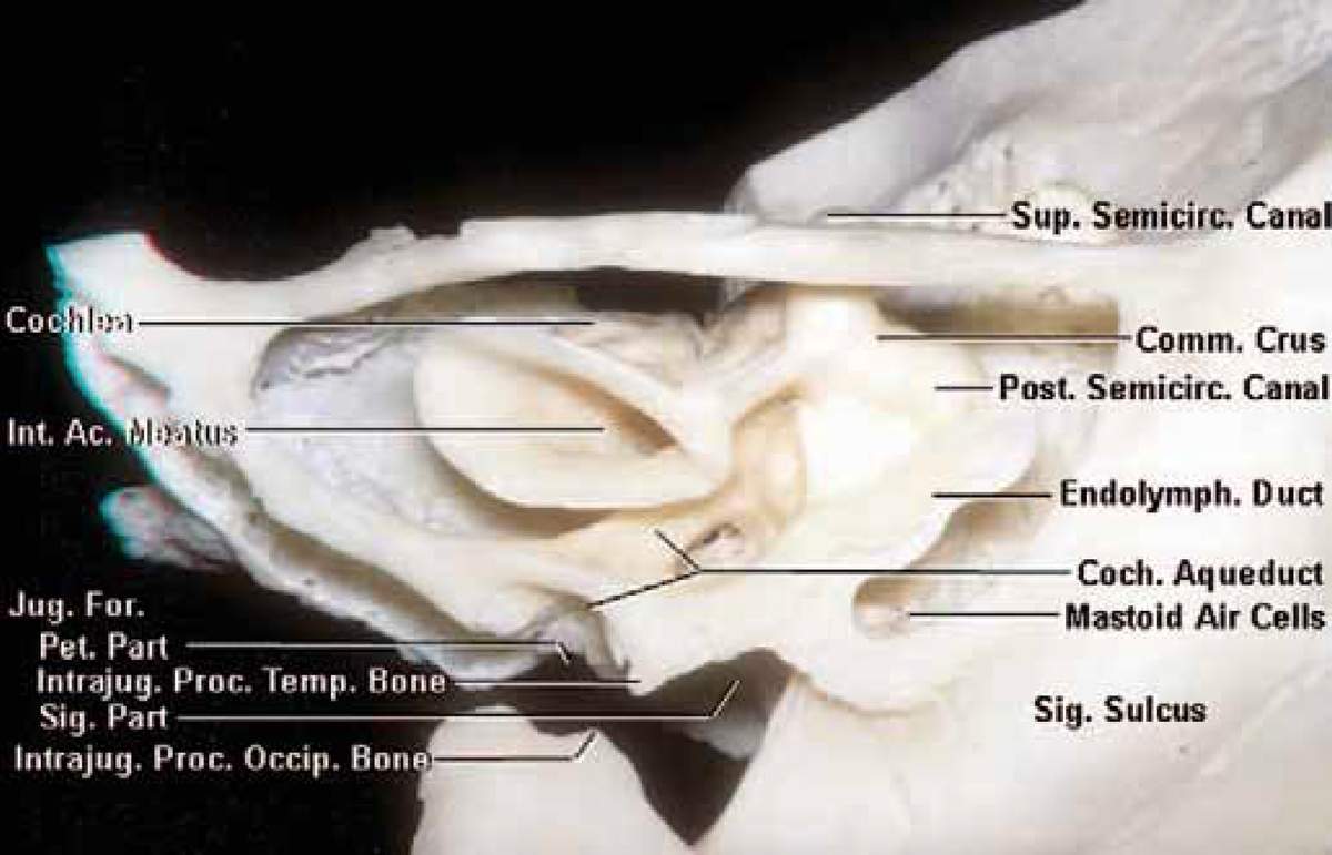
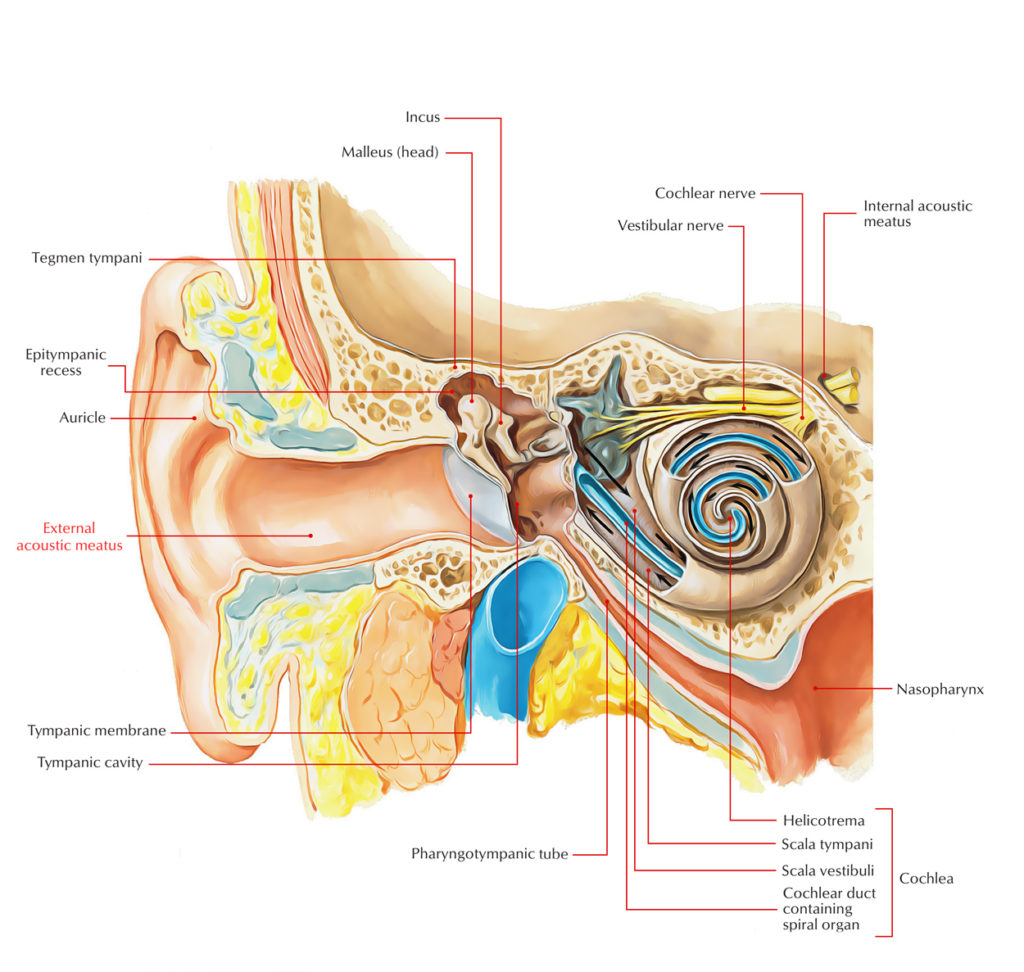
The internal auditory canal (IAC), also referred to as the internal acoustic meatus lies in the temporal bone and exists between the inner ear and posterior cranial fossa. It includes the vestibulocochlear nerve (CN VIII), facial nerve (CN VII), the labyrinthine artery, and the vestibular ganglion.
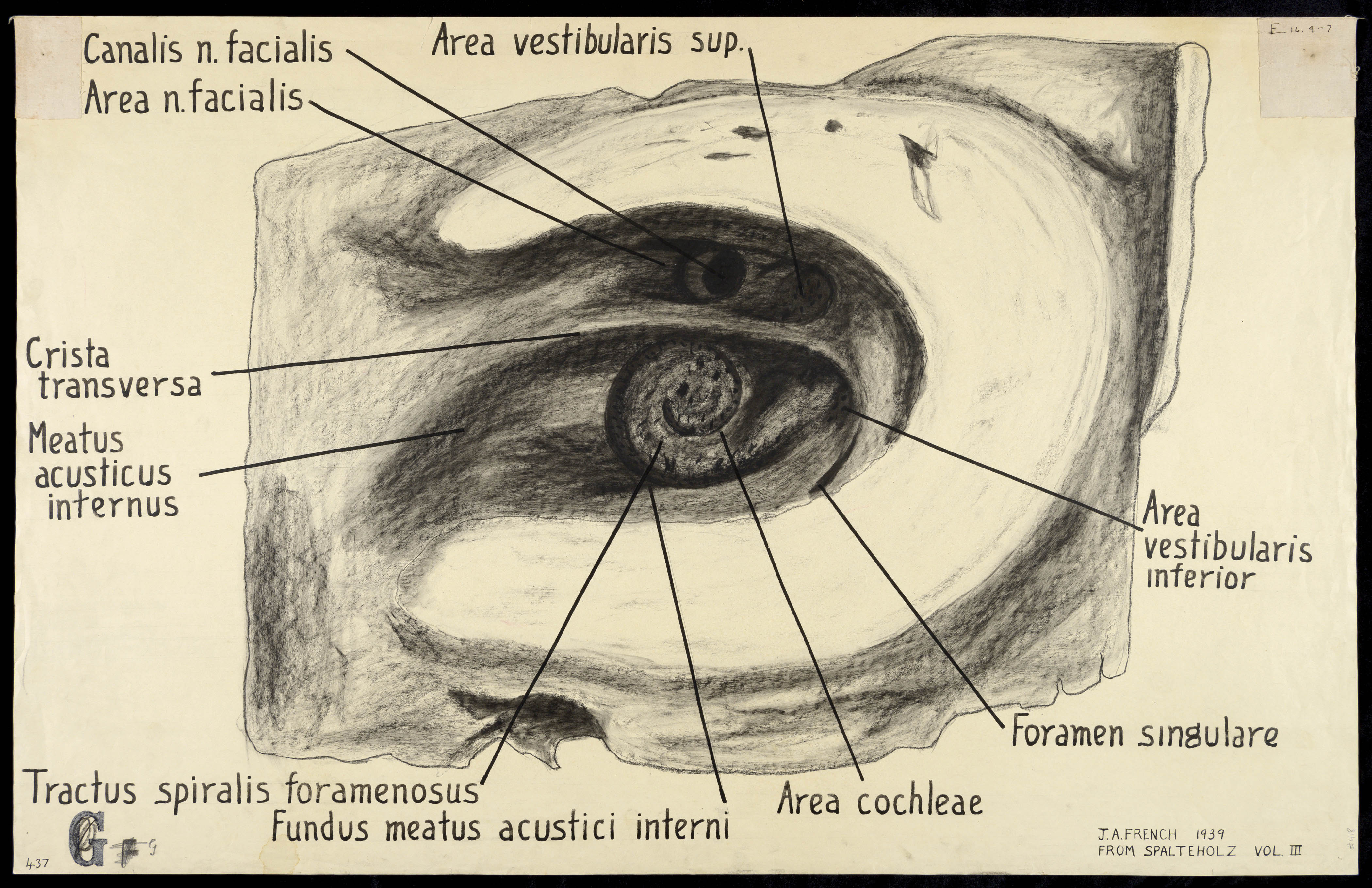
Canalis n. facialis, Area n. facialis, Crista transversa, Meatus acusticus internus...
Definition noun A short, narrow passageway through the temporal bone of the skull where the vestibular nerve and cochlear nerve pass through to reach the brainstem from the inner ear. Supplement In humans, the internal acoustic meatus is about 1-2 cm in length.

Odyoloji Okuyoruz Biz
The internal acoustic meatus was evaluated in 97 temporal bone specimens, half of which were radiographed in different projections.. Die Varianten des Meatus acusticus internus. Radiol. Diagn. 7 (1966), 141. PubMed. Google Scholar. 5. Camp J., Cilley.: The significance of asymmetry of the pori acustici as an aid in diagnosis of eighth nerve.
HISTOLOGI TELINGA
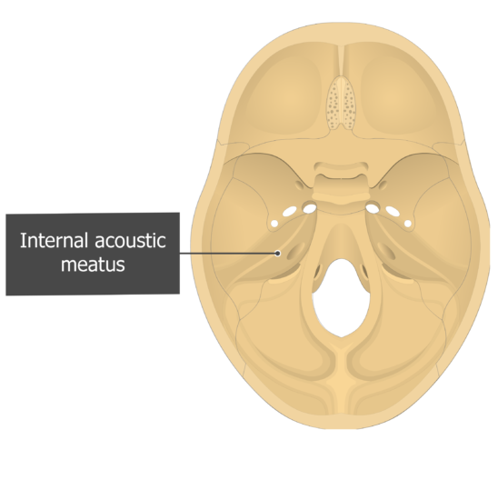
Internal acoustic meatus refers to a small bony foramen situated on the posterior surface of the petrous part of temporal bone, inside the posterior cranial fossa. It allows for the passage of three important structures, namely the vestibulocochlear nerve, facial nerve and the labyrinthine artery.
Cerebellopontine Angle and Cranial Nerves The Neurosurgical Atlas, by Aaron CohenGadol, M.D.
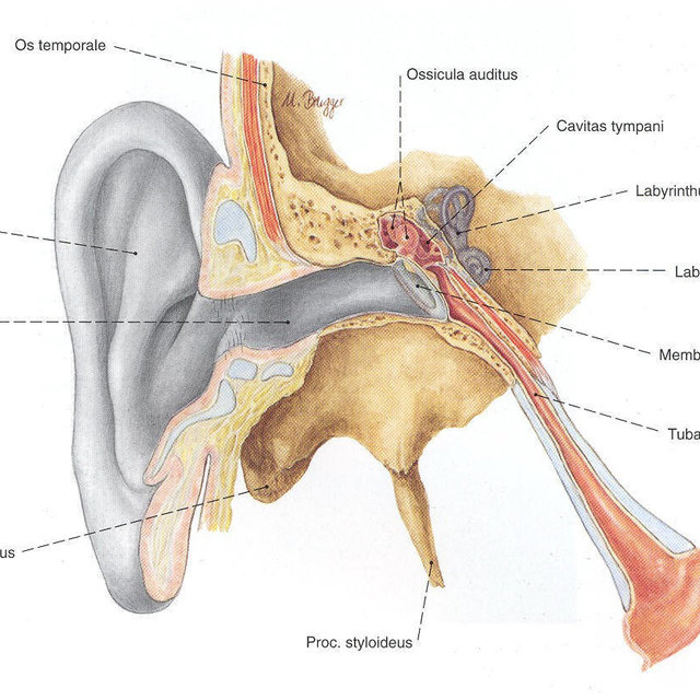
The internal auditory meatus (also meatus acusticus internus, internal acoustic meatus, internal auditory canal, or internal acoustic canal) is a canal within the petrous part of the temporal bone of the skull between the posterior cranial fossa and the inner ear . Structure
External Ear , Auricle and External acoustic meatus , Anatomy QA
A trumpeted internal acoustic meatus (IAM) is an indirect sign of a vestibular schwannoma and is useful in helping to differentiate between one and other cerebellopontine angle entities, especially from a meningioma which typically does not extend into the meatus and is more often associated with hyperostosis 1.. It is characterized by widening of the medial end of the internal acoustic canal.
Oído medio Anatomía, partes, funciones Kenhub
The internal auditory canal (IAC), also referred to as the internal acoustic meatus lies in the temporal bone and exists between the inner ear and posterior cranial fossa. It includes the vestibulocochlear nerve (CN VIII), facial nerve (CN VII), the labyrinthine artery, and the vestibular ganglion..
external acoustic meatus bone location
The internal auditory meatus (IAM) is a small, bony canal located within the petrous portion of the temporal bone of the skull. It serves as an important pathway for various cranial nerves and blood vessels to pass through the inner ear.
Internal Acoustic Meatus
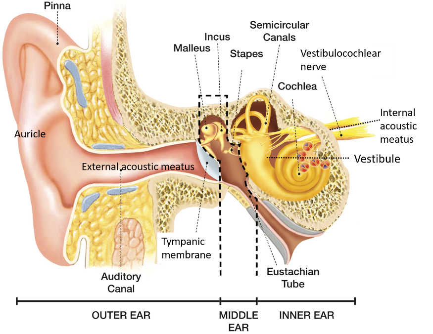
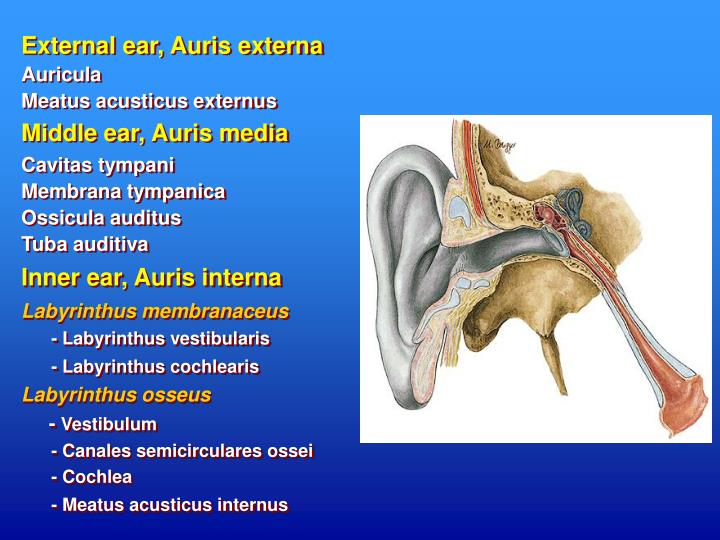
Internal ear This mixture of bones, nerves, vessels, membranes, and muscles that make up the ear will be described in this article. Contents External ear Auricle External acoustic meatus Tympanic membrane Muscles of the external ear Vasculature of the external ear Innervation of the external ear Middle ear Tympanic cavity Auditory ossicles
1 Semischematic overview of the outer (Auricula and Meatus acusticus... Download Scientific
The internal acoustic canal (IAC) , also known as the internal auditory canal or meatus (IAM), is a bony canal within the petrous portion of the temporal bone that transmits nerves and vessels from within the posterior cranial fossa to the auditory and vestibular apparatus. Gross anatomy
Pterygopalatine Fossa Anatomy and Contents Kenhub
The porus acusticus internus (plural: pori acustici interni), often merely referred to as porus acusticus, is the medial opening of the internal acoustic canal through which the facial nerve , vestibulocochlear nerve and labyrinthine artery pass 1 .
PPT Anatomy of the Ear PowerPoint Presentation ID6102402
The influences of porus acusticus internus on ethnicity and importance in preoperative and intraoperative approaches. R. Şekerci Eren Ogut N. Keles-Celik. Medicine.. Amac: Bu calisma 18-60 yas arasi saglikli Turk populasyonunda meatus acusticus internus morfometrisini belirlemek amaclandi, yurutulen bu calisma retrospektif bir calismadir.
External Auditory Meatus/Acoustic Meatus Earth's Lab
MRI is firmly established as an essential modality in the imaging of the temporal bone and lateral skull base. It is used to evaluate normal anatomic structures, evaluate for vestibular schwannomas, assess for inflammatory and/or infectious processes, and detect residual and/or recurrent cholesteatoma. It is also extensively used in pre- and postoperative evaluations, particularly in patients.

Meatus acusticus internus Ars Neurochirurgica
Definition The internal acoustic opening is a large orifice near the center of posterior surface of petrous part, its size varies considerably; its margins are smooth and rounded, and it leads into a short canal, the internal acoustic meatus, about 1 cm. in length, which runs lateralward.
