
Pin by Kaela WrightCook on RN Eye anatomy diagram, Human eye diagram, Diagram of the eye
Reviewed/Revised Mar 2022 | Modified Sep 2022 VIEW PROFESSIONAL VERSION The structures and functions of the eyes are complex. Each eye constantly adjusts the amount of light it lets in, focuses on objects near and far, and produces continuous images that are instantly transmitted to the brain.
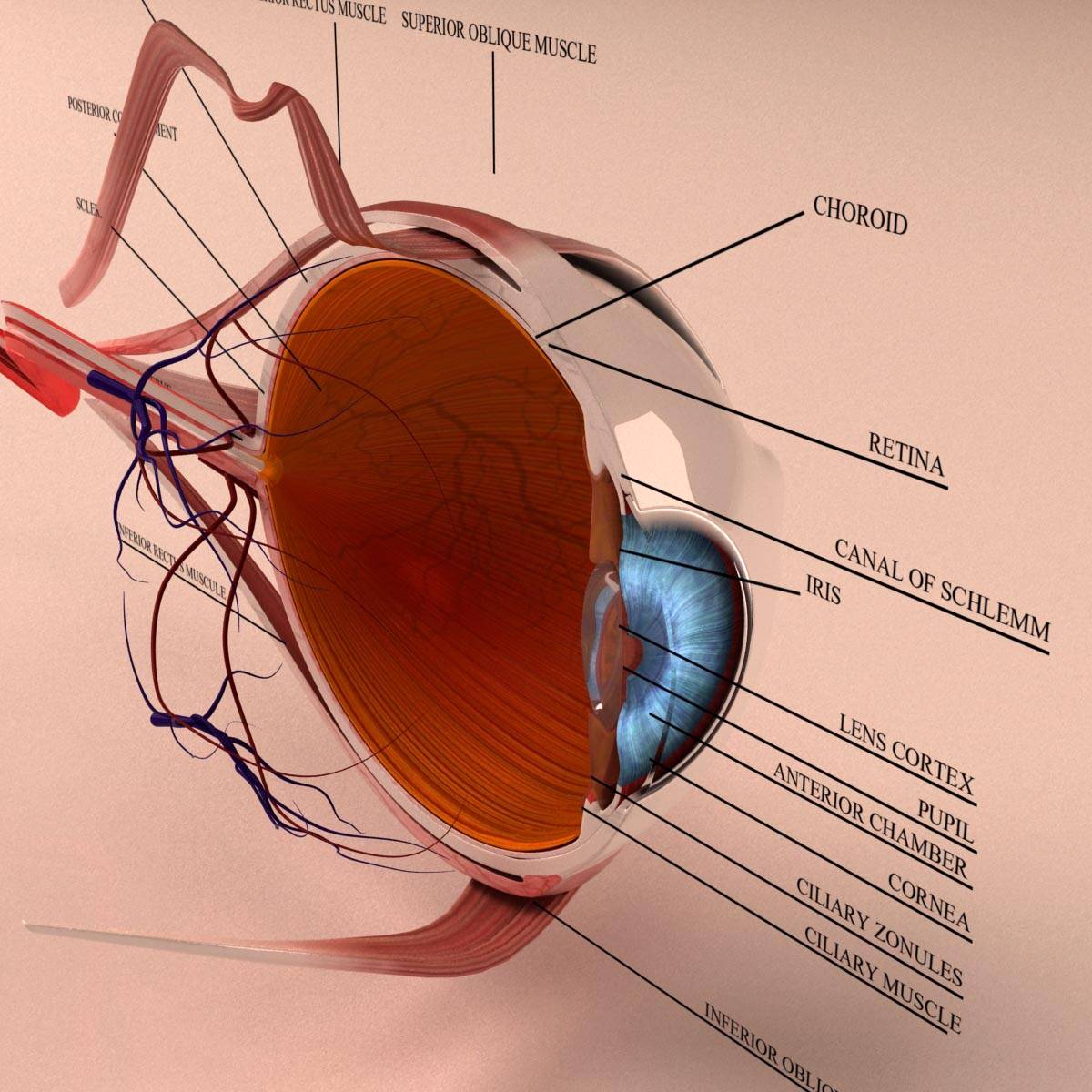
Anatomy Human Eye Cross Section 3D Model Kezan's Portfolio
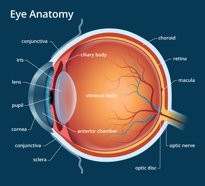
View All. Cornea. Pupil. Iris. Crystalline Lens. Aqueous Humor. The human eye is an organ that detects light and sends signals along the optic nerve to the brain. Perhaps one of the most complex organs of the body, the eye is made up of several parts—and each individual part contributes to your ability to see.

How Your Eye Works Optometry in Denver Optical Masters
Inner layer Blood supply of the eye Nerves of the eye Sources + Show all Bones of the orbit The bony orbit is made out of seven bones, which include the maxilla, zygomatic bone, frontal bone, ethmoid bone, lacrimal bone, sphenoid bone and palatine bone.
Eye Anatomical Chart
Structure A detailed depiction of eye using a 3D medical illustration MRI scan of the human eye Humans have two eyes, situated on the left and the right of the face. The eyes sit in bony cavities called the orbits, in the skull. There are six extraocular muscles that control eye movements.
/GettyImages-1128675065-e4bac15b0f39449dba31f25f1020bc8a.jpg)
An Overview of Eye Anatomy
Your eye is a slightly asymmetrical globe, about an inch in diameter. The front part (what you see in the mirror) includes: Iris: the colored part Cornea: a clear dome over the iris Pupil: the.
Vision and Eye Diagram How We See
The coloured part of your eye is called the iris. The iris is made up of muscle fibres which help to control the size of the pupil. The pupil is not an actual structure but the circular opening in the middle of the iris. The pupil appears as the dark central part of the eye. The pupil can change size (through changes in the iris) in order to.
Human Eye Anatomy, parts and structure Online Biology Notes
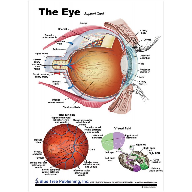
This popular chart of The Eye has illustrations by award winning medical illustrator Keith Kasnot. The chart covers general anatomy of the eye with colorful detailed renderings all fully labeled. Includes the following images: the outer eye as we see it with all parts labeled lateral view of the eyeball in the skull top view of the eyeball in the skull diagram of the visual field large central.
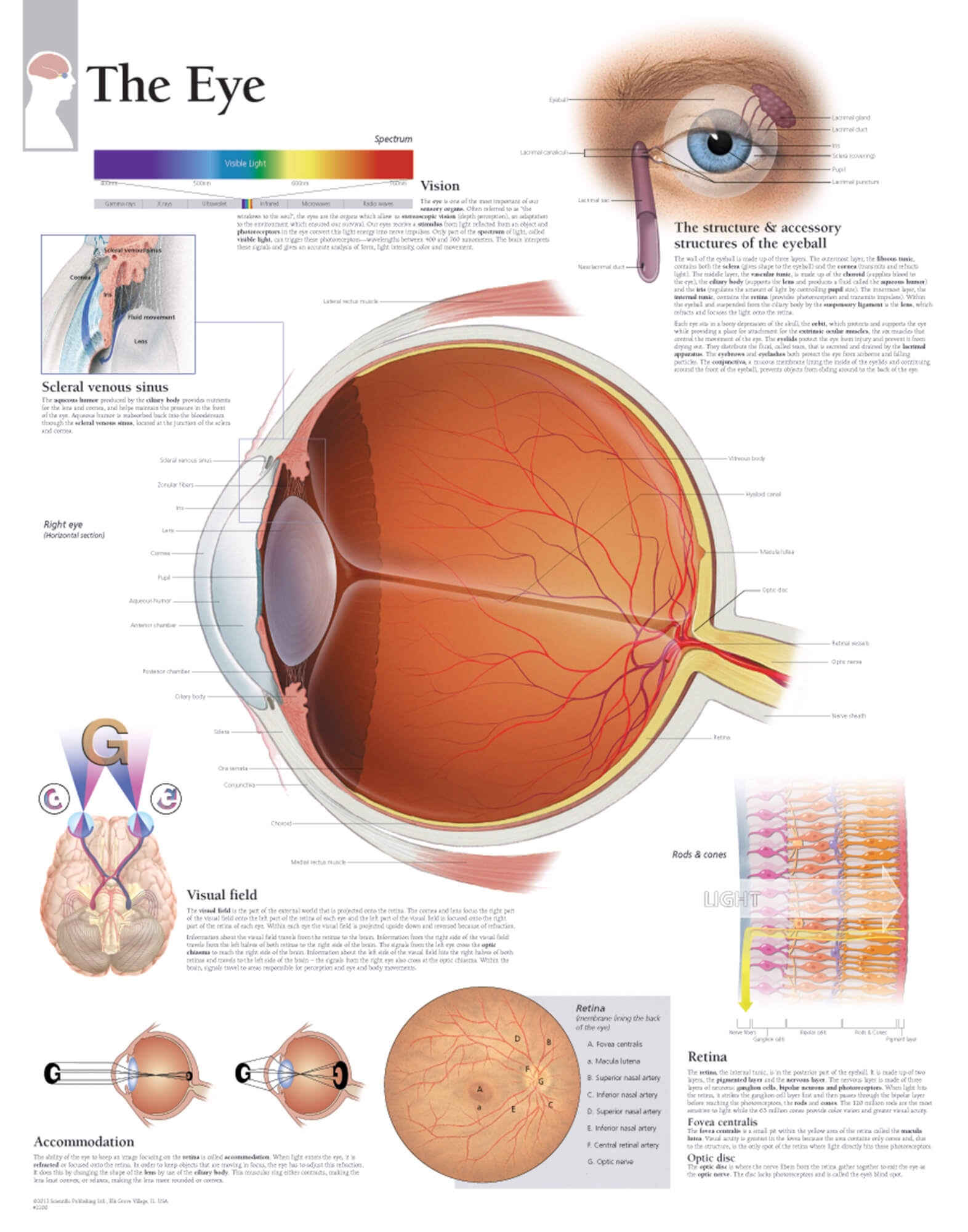
The Eye Scientific Publishing
Iris: regulates the amount of light that enters your eye. It forms the coloured, visible part of your eye in front of the lens. Light enters through a central opening called the pupil. Pupil: the circular opening in the centre of the iris through which light passes into the lens of the eye. The iris controls widening and narrowing (dilation and.

OUR EYES WORK LIKE CAMERA’S! Discovery Eye Foundation
The main blood supply of the eye arises from the ophthalmic artery, which gives off orbital and optical group branches. Innervation of the eyeball and surrounding structures is provided by the optic, oculomotor, trochlear, abducens and trigeminal cranial nerves. This article covers the anatomy, function and clinical relevance of the vessels and.
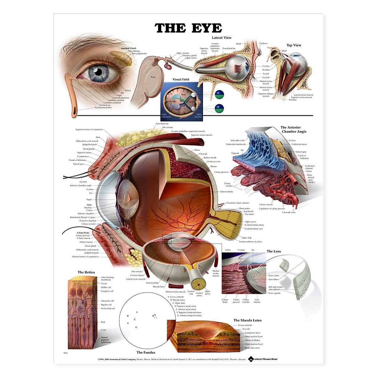
The Eye Anatomical Chart 20'' x 26''
Eye anatomy: A closer look at the parts of the eye By Liz Segre Understanding how vision works When surveyed about the five senses — sight, hearing, taste, smell and touch — people consistently report that their eyesight is the mode of perception they value (and fear losing) most.

Human Eye Anatomy, Structure and Function
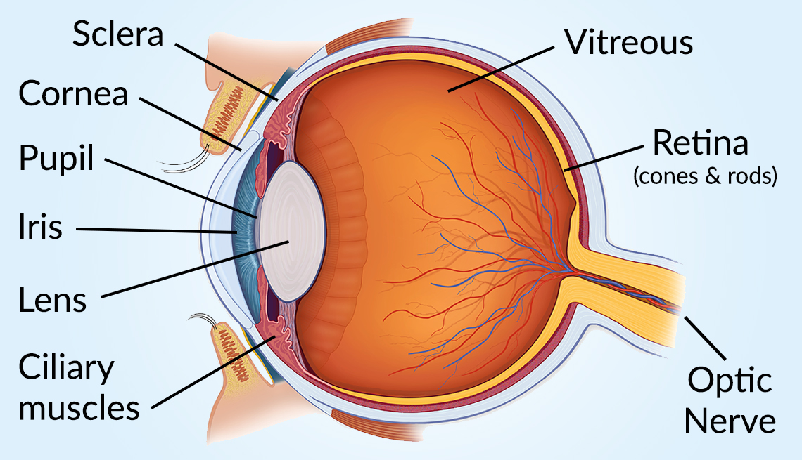
Eyes are approximately one inch in diameter. Pads of fat and the surrounding bones of the skull protect them. The eye has several major components: the cornea, pupil, lens, iris, retina, and sclera.
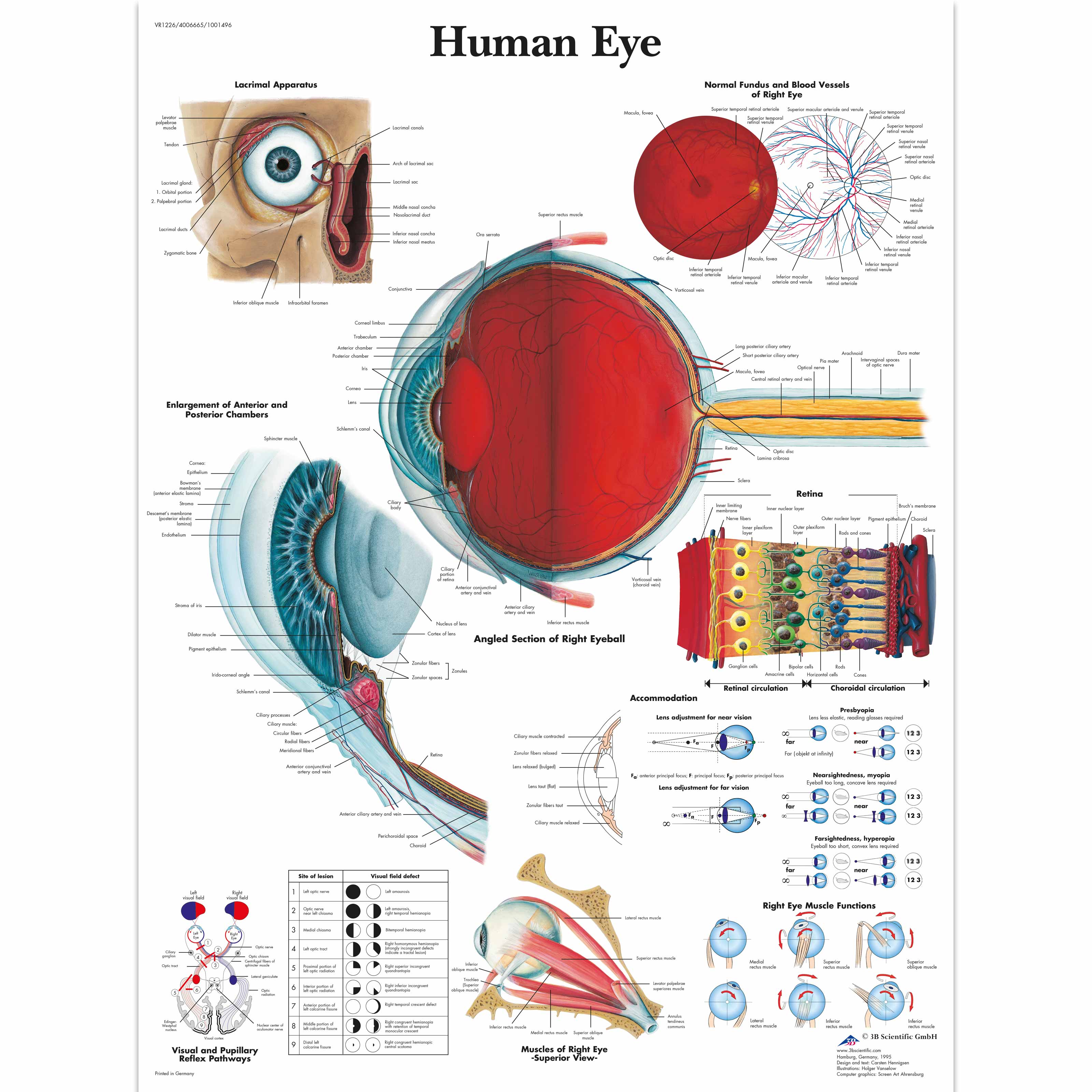
Human Eye Chart 1001496 3B Scientific VR1226L Ophthalmology charts and posters
How Do the Eyes Work? Eye Anatomy (16 Parts of the Eye & What They Do) Summary How Do the Eyes Work? Light is reflected when you focus on an object and enters the eye through the cornea. As the light passes through, the dome-shaped nature of the cornea bends light, enabling the eye to focus on fine details.

Eye Anatomy, Functions, Diseases, Diagnosis, Tips For Good Eye Sight LeoGenic Healthcare Pvt Ltd
The surface of the eye and the inner surface of the eyelids are covered with a clear membrane called the conjunctiva. The layers of the tear film keep the front of the eye lubricated. Tears lubricate the eye and are made up of three layers. These three layers together are called the tear film. The mucous layer is made by the conjunctiva.

Structure Of Human Eye Human Sense Organs The Five Senses A comprehensive guide to human
The iris is a flat, thin, ring-shaped structure sticking into the anterior chamber. This is the part that identifies a person's eye colour. The iris contains both circular muscles going around the pupil and radial muscles radiating toward the pupil. When the circular muscles contract, they make the pupil smaller.
Eye Anatomy Chart B
Eye anatomy: A closer look at the parts of the eye. By Liz Segre. When surveyed about the five senses — sight, hearing, taste, smell and touch — people consistently report that their eyesight is the mode of perception they value (and fear losing) most. Despite this, many people don't have a good understanding of the anatomy of the eye, how.

Eye Pain Pain Behind, Sharp Pain, Eye Pain With Headache Causes
Anatomy of the Human Eye. Eyes are one of the most important organs of the body. A healthy pair of eyes means a clear vision, which plays a major role in day-to-day life and quality of experiences.
