9. Muscles of the Head Musculoskeletal Key
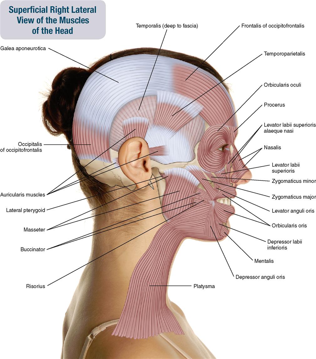
The facial muscles can be split into three groups: orbital, nasal and oral. Orbital Group. Schematic of head and neck muscles.: Locations of facial muscles noted.. The risorius muscle is lateral to the orbicularis oris and inserts into the angle of the mouth. When innervated, the risorius pulls the mouth back mimicking a smile, but does not.
Facial Muscles and Expressions Classic Human Anatomy in Motion The
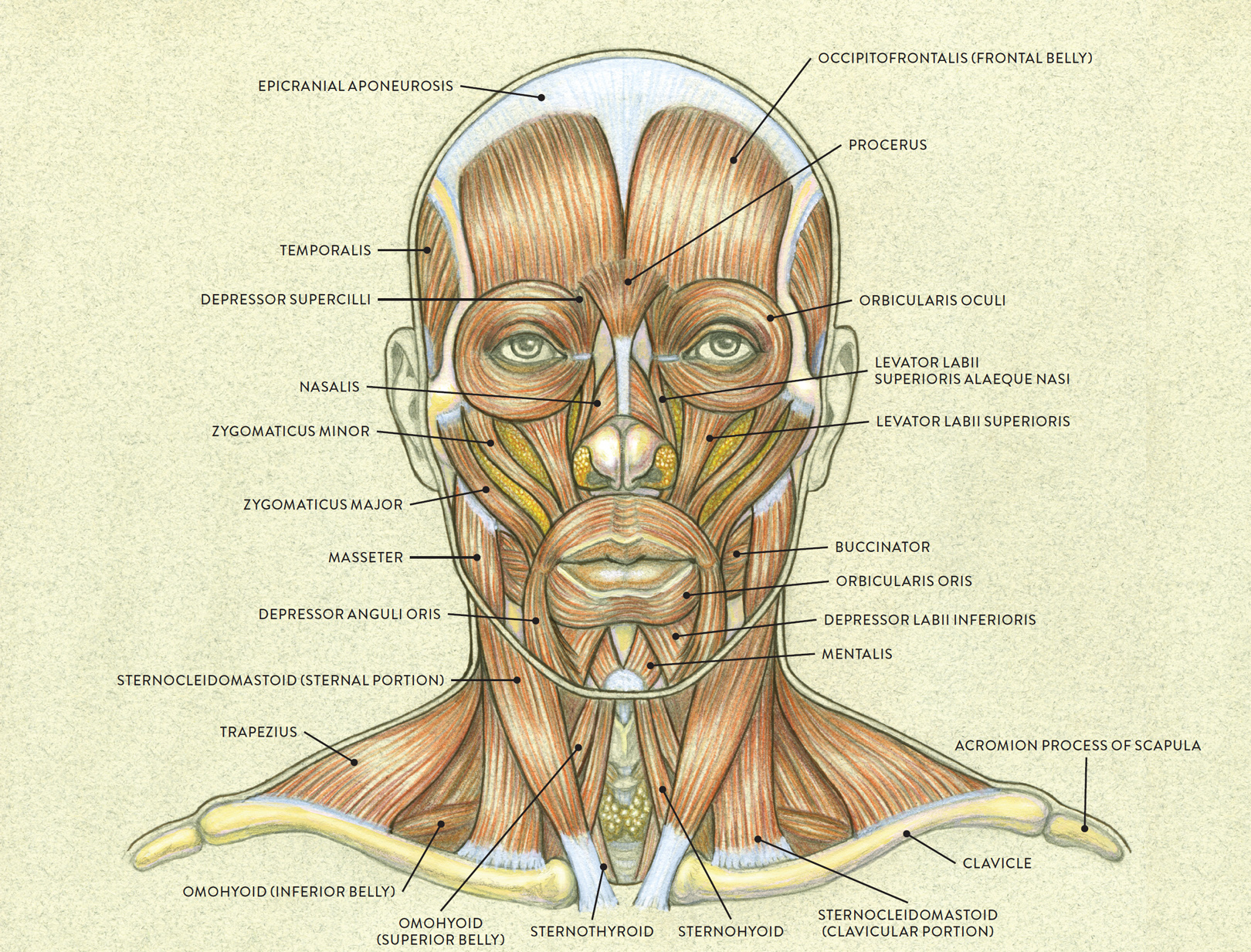
The facial muscles are just under the skin ( subcutaneous) muscles that control facial expression. They generally originate from the surface of the skull bone (rarely the fascia), and insert on the skin of the face. When they contract, the skin moves. These muscles also cause wrinkles at right angles to the muscles' action line.

Muscles of the head (left lateral view) PurposeGames
The facial muscles are positioned around facial openings (mouth, eye, nose and ear) or stretch across the skull and neck. Thus, these muscles are categorized into several groups; Muscles of the mouth (buccolabial group) Muscles of the nose (nasal group) Muscles of the cranium and neck Muscles of the external ear (auricular group)

Musculos Da Face Lateral
1.10.1 Aging Process of the Facial Tissue. The anatomical structures of the face related to aging comprise of the facial bone, fat tissue, fibrous connective tissue, and facial muscles. The bony tissue is a structure that forms the basic frame of the face and bone remodeling goes throughout lifelong period.

6 Lateral view of the Facial Muscles Download Scientific Diagram
Facial Muscles: Anatomy The facial muscles (also called mimetic muscles) control facial expression and are supplied by the facial nerve. Most of them originate from the skull and attach to the skin around the facial openings, which serve as a method to group or classify them.

Muscles of the face and neck lateral view (2) Diagram Quizlet
The muscles of facial expression are innervated by the facial nerve (cranial nerve VII), and the muscles of mastication are innervated by the mandibular division of the trigeminal nerve (cranial nerve V3). [1]
Zygomaticus major hires stock photography and images Alamy
The neck muscles, including the sternocleidomastoid and the trapezius, are responsible for the gross motor movement in the muscular system of the head and neck. They move the head in every direction, pulling the skull and jaw towards the shoulders, spine, and scapula. Working in pairs on the left and right sides of the body, these muscles.

Dentistry lectures for MFDS/MJDF/NBDE/ORE A Note on Muscles of the
Structure and Function The anatomy of the face can divide into three main regions: upper face, middle face, and lower face. The entire face is covered by skin superficially, while the deep anatomy contains muscles, fat pads, nerves, vessels, and bones. Upper Face

Body muscle anatomy, Head muscles, Muscle
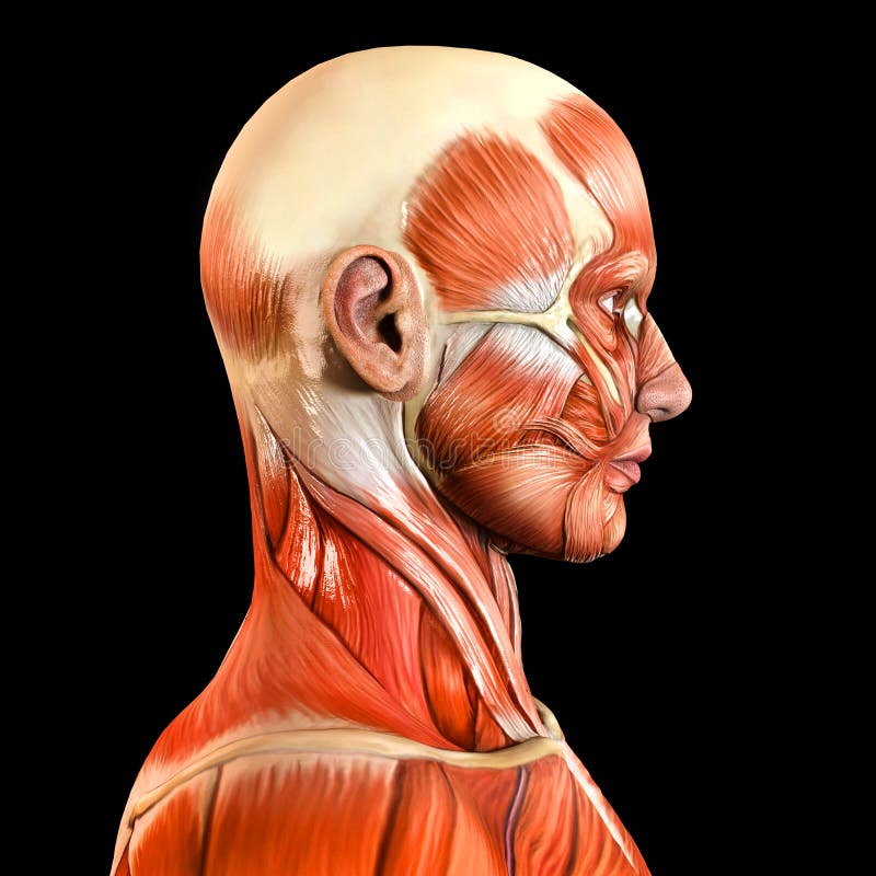
Found situated around openings like the mouth, eyes and nose or stretched across the skull and neck, the facial muscles are a group of around 20 skeletal muscles which lie underneath the facial skin. The majority originate from the skull or fibrous structures, and connect to the skin through an elastic tendon.

Face And Neck Muscle Diagram / Facial Muscles Images Stock Photos
The superficial motor nerves to the muscles of facial expression from the facial nerve (temporal, zygomatic, buccal, mandibular, cervical branches, and the posterior auricular nerve) are described. The sensory nerves to the face (branches of each of the three divisions of the trigeminal nerve or cervical nerves) are delineated.

Axial Muscles of the Head, Neck, and Back · Anatomy and Physiology
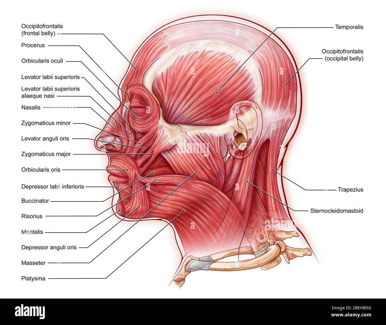
Lateral View of Skull. A view of the lateral skull is dominated by the large, rounded cranium above and the upper and lower jaws with their teeth below.. The origins of the muscles of facial expression are on the surface of the skull. The insertions of these muscles have fibers intertwined with connective tissue and the dermis of the skin.

Lateral Superficial Facial Muscles
Definition. Elevates mandible as in closing mouth, assists in side-to-side movement of mandible, and protracts (protrudes) mandible. Location. Term. Sternocleidomastoid Muscle. Definition. Contraction of both muscle flexes the cervical part of the vertebral column and draws the head forward; contraction of one muscle rotates the face toward.
Lateral Side Facial Face Muscles Stock Photo Image 48360396
Muscles of Facial Expression, Lateral View 5.0 (1 review) Flashcards Learn Test Match Flashcards Learn Test Match Created by Hildhanga Terms in this set (10) Term Masseter Location Term Levator labii superioris Location Term Platysma (cut and reflected) Location Term Depressor anguli oris Location Term Buccinator Location Term Orbicularis oculi

Muscle of the head, lateral view, illustration Stock Image C039
The facial muscles can broadly be categorised into three groups - orbital, nasal and oral. In this article, we shall look at the anatomy of the muscles of facial expression - their attachments, actions and clinical relevance. Fig 1 - Innervation to the muscles of facial expression via the facial nerve (CN VII) Orbital Group

The Muscles of the Head allowing face mimics and mastication
The SMAS consists of three distinct layers: (1) a fascial layer superficial to the muscles, (2) a layer intimately associated with the facial m., and (3) a deep layer extensively attached to the periosteum of facial bones (Fig. 2.2 ). Fig. 2.2 Photographs showing the dissection of SMAS and subSMAS fat.

Face Muscles. FaceMuscle. LateralFaceMuscles. Anatomy.
The facial muscles (also known as the muscles of facial expression) are situated within the subcutaneous tissue of the face and responsible for the movements of skin folds, providing different facial expressions.. The facial muscles originate from bones of the facial skeleton (viscerocranium) and insert into the skin.; The facial muscles are mostly grouped around the natural orifices of the.
