Human Body Mrs. Willis 7th Life Science
Muscular. The primary job of muscles is to move the bones of the skeleton, but muscles also enable the heart to beat and constitute the walls of other vital hollow organs. Skeletal muscle: This.

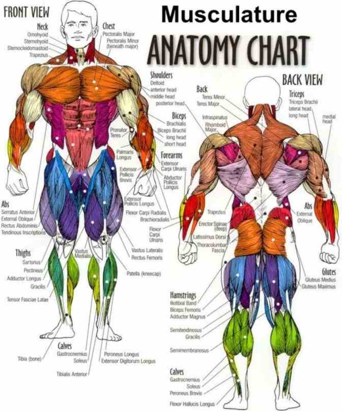
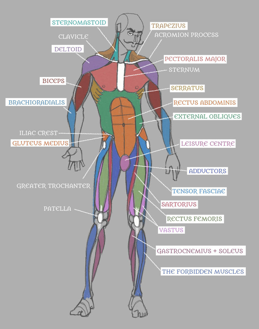
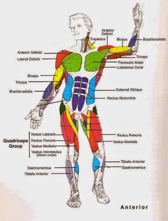
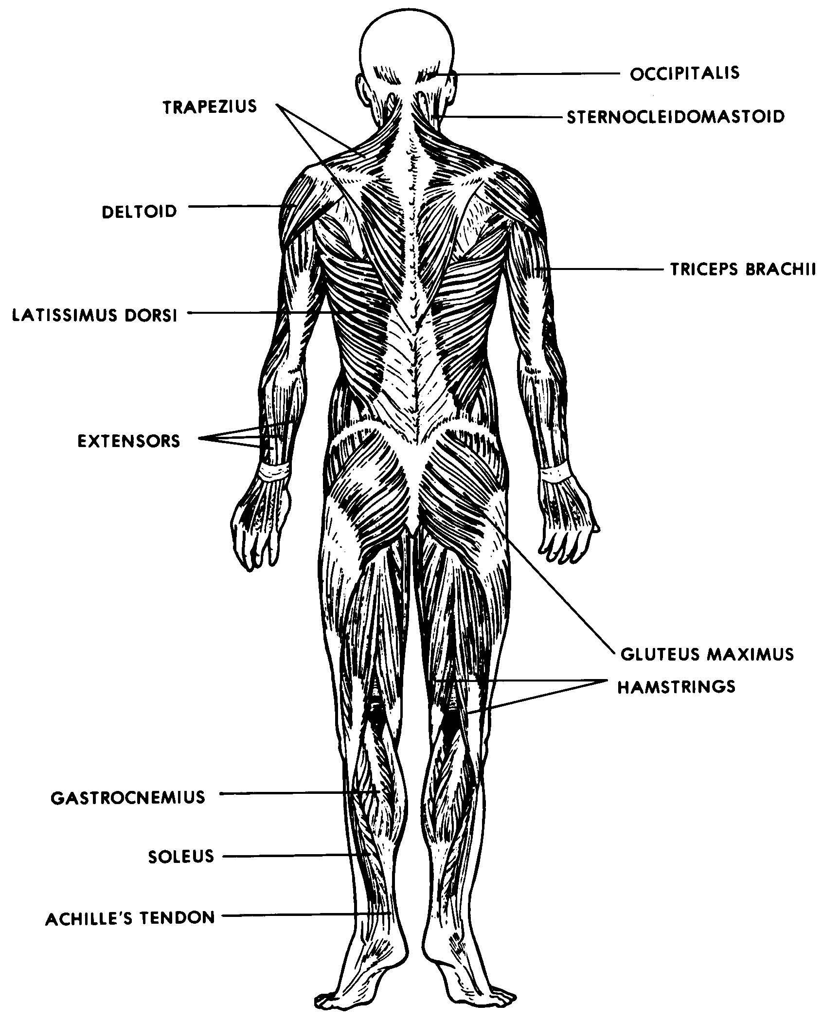
Labeled Muscle Diagram Chart Free Download
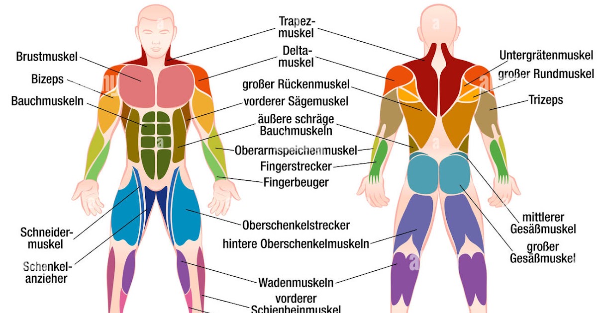
Muscle anatomy quiz for anatomy and physiology! When you are taking anatomy and physiology you will be required to identify major muscles in the human body. This quiz requires labeling, so it will test your knowledge on how to identify these muscles (latissimus dorsi, trapezius, deltoid, biceps brachii, triceps brachii, brachioradialis, pectoralis major, serratus anterior, rectus abdominis, etc.).
34 Label Muscular System Worksheet Labels Design Ideas 2020
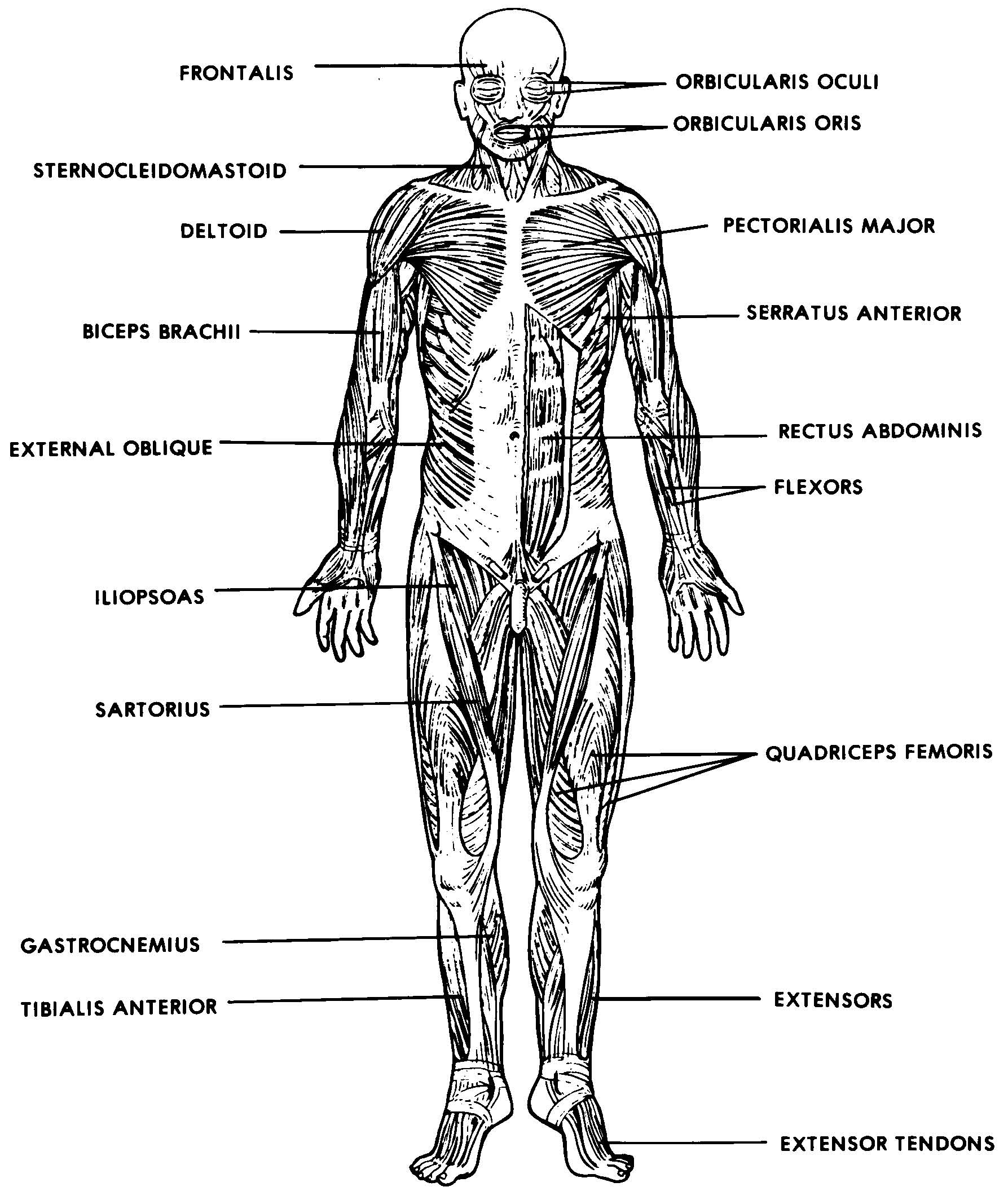
Forearm muscle anatomy. The forearm is a region of the upper extremity extending from the wrist to the elbow joint. It is split into two compartments: anterior and posterior. In the posterior compartment, which is what we'll be focusing on in this article, we find the extensor muscles. These muscles of the forearm are responsible for.

FileMuscle posterior labeled.png Wikipedia
Human body muscle diagrams. Muscle diagrams are a great way to get an overview of all of the muscles within a body region. Studying these is an ideal first step before moving onto the more advanced practices of muscle labeling and quizzes. If you're looking for a speedy way to learn muscle anatomy, look no further than our anatomy crash courses .
Muscle Labeling Diagram Human Body Anatomy
The muscular system is made up of three types of muscle tissue: skeletal, cardiac and smooth. In this section, we'll focus on the skeletal muscles of the body involved in voluntary movement and maintaining posture. They are attached to bones via tendons and contract to cause movement at the joints. Learn more about the anatomy and functions of.
Simple Human Muscles Diagram / Learn All Muscles With Quizzes And
The revolutionary breakthrough in understanding the meaning of our human conditioned lives. The desperately needed scientific breakthrough that finally solves the meaning of life.

Diagram Of Muscles In The Body Amazon Com Human Body Muscle Anatomy
Chapter 8: Muscular System. This chapter is divided into three main sections: muscle basics and cellular components, naming of the muscles, and cat muscles with dissection.. Interactive Diagrams: Head | Back | Chest | Legs (anterior). More Labeling - Head | Full Body | Full Body Side. Part 3: Cat Muscles and Dissection.
Simple Human Muscles Diagram Major Muscles Of The Human Body For Kids
Inner hip & gluteal muscles. Anterior, medical and posterior thigh muscles. Anterior, lateral and posterior leg muscles. Dorsal and plantar foot muscles. This eBook contains high-quality illustrations and validated information about each muscle. It is available for free. Download free PDF (8.5MB) Get for free on iBooks.

Human Muscles Diagram Muscle Diagram Anatomy System Human Body Images
Human muscle system, the muscles of the human body that work the skeletal system, that are under voluntary control, and that are concerned with movement, posture, and balance. Broadly considered, human muscle—like the muscles of all vertebrates—is often divided into striated muscle, smooth muscle, and cardiac muscle.
Labeled Body Muscle Diagram
Don't swipe away. Massive discounts on our products here - up to 90% off! Come and check all categories at a surprisingly low price, you'd never want to miss it.
Human Anatomy Muscles with Labels! by Pseudolonewolf on DeviantArt
externus. outside. EXternal. internus. inside. INternal. Table 11.2. Anatomists name the skeletal muscles according to a number of criteria, each of which describes the muscle in some way. These include naming the muscle after its shape, its size compared to other muscles in the area, its location in the body or the location of its attachments.

Label Muscles Worksheet Human muscle anatomy, Muscle anatomy, Human
The muscular system is responsible for the movement of the human body. Attached to the bones of the skeletal system are about 700 named muscles that make up roughly half of a person's body weight. Each of these muscles is a discrete organ constructed of skeletal muscle tissue, blood vessels, tendons, and nerves.

Unlabelled Diagram Of Muscles In The Body / Muscles Of The Body Blank
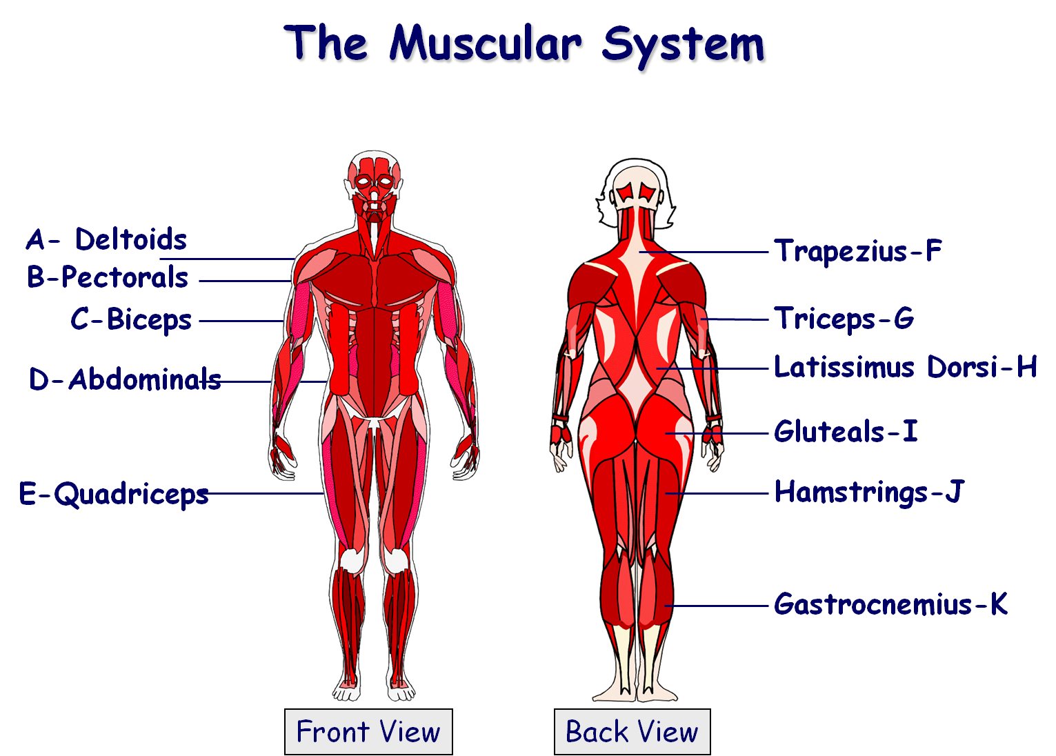
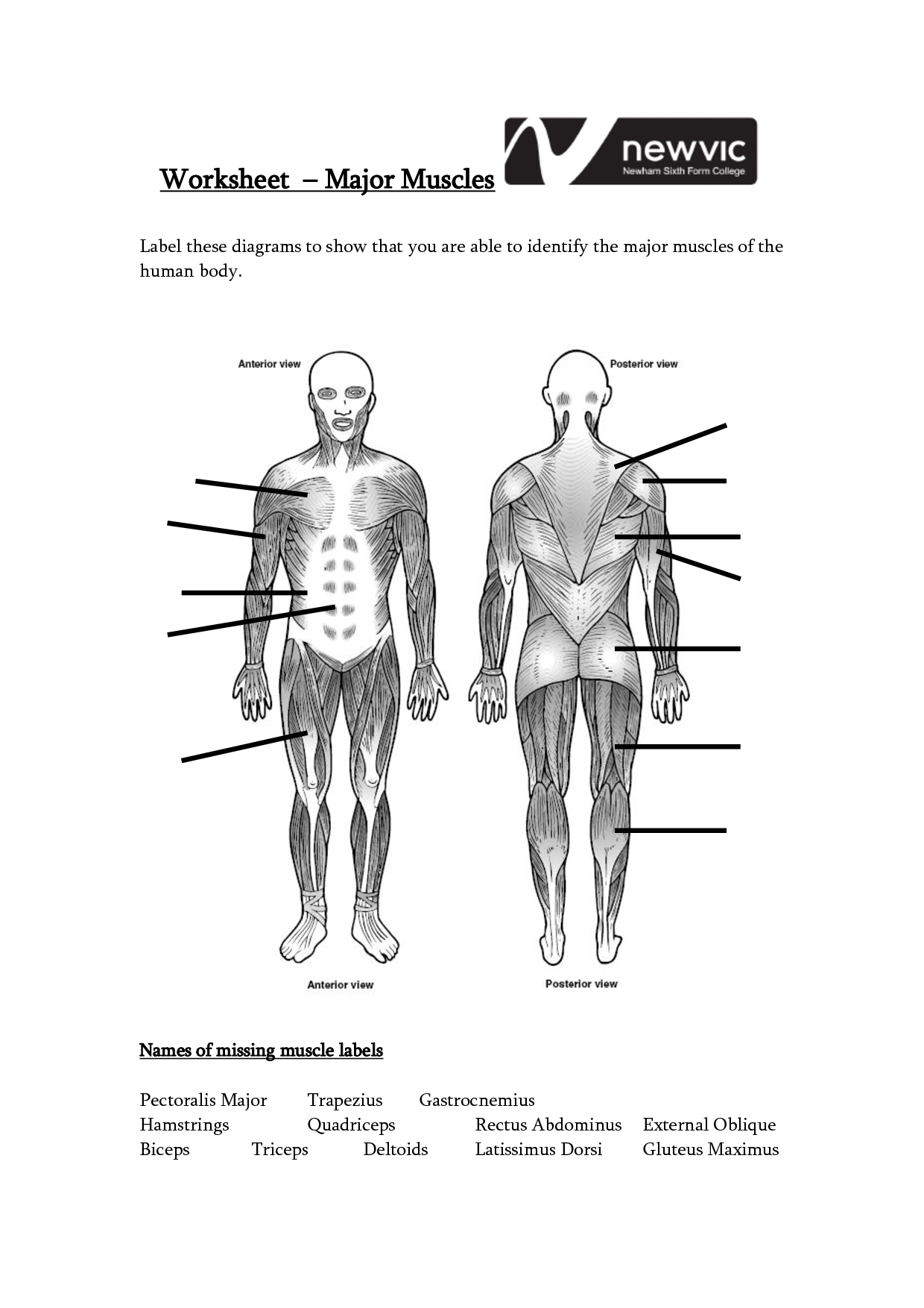
Use this set of muscular system body diagram to help your children learn all of the major skeletal muscle groups of the human body. Children are required to correctly allocate each muscle name to its corresponding label. This informative resource is perfect for teaching about muscles (KS2 level). It has 10 of the more widely-known muscles, including abdominals, pectorals and hamstrings.
Labeled Muscle System Holland Teenpornclips
Each skeletal muscle is an organ that consists of various integrated tissues. These tissues include the skeletal muscle fibers, blood vessels, nerve fibers, and connective tissue. Each skeletal muscle has three layers of connective tissue (called "mysia") that enclose it and provide structure to the muscle as a whole, and also.
Printable Muscle Labeling Worksheet
Start studying Anatomy & Physiology - Muscle labeling. Learn vocabulary, terms, and more with flashcards, games, and other study tools.

Back Muscles Anatomy Labeled Upper Back Anatomy and physiology
This muscular system label activity is a fun and engaging way for learners to review and extend their knowledge.Muscle Labelling would be a great exercise for a Science or STEM lesson. According to the Australian Curriculum, it isn't essential for primary level children to learn about the muscles of the human body. That being said, this worksheet would still make a good practice for laying.
